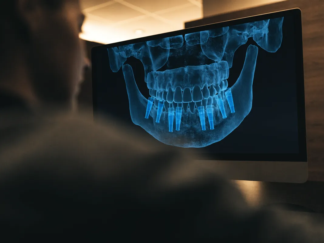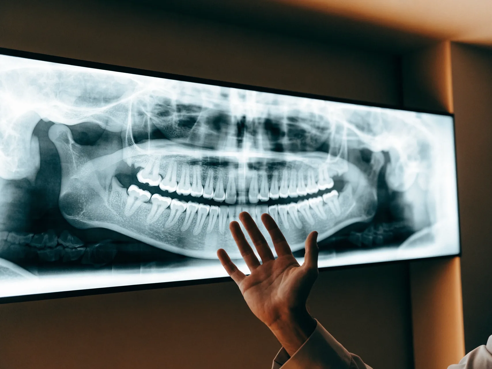
A high-quality panoramic X-ray is the foundation of any useful remote dental consultation. Here is exactly how to obtain one (free or low-cost), capture it correctly, and send it to us so we can give you accurate advice.
A useful remote enquiry depends heavily on the quality and date of the records. A panoramic X-ray may help a named clinician discuss possible options, but it does not replace examination or any imaging that clinician considers necessary. This guide explains how to obtain and transmit an X-ray securely and how to recognise a provisional plan.
What We Need
The single most useful image is a panoramic dental X-ray — also called an OPG (Orthopantomogram) or DPT (Dental Panoramic Tomogram). This shows your entire upper and lower jaws in one image, including:
- All visible teeth
- Tooth roots and surrounding bone
- Sinus boundaries (upper jaw)
- Inferior alveolar nerve (lower jaw)
- Any existing fillings, root canals, crowns, or implants
- Areas of bone loss or pathology
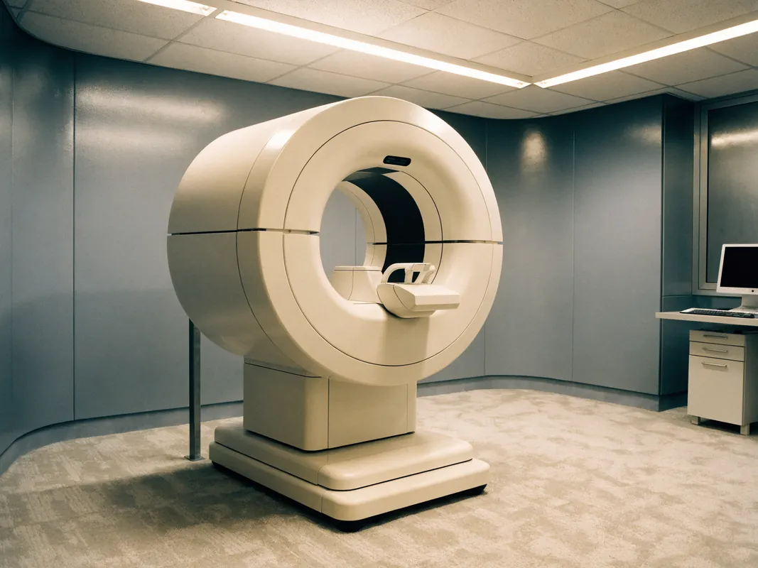
A panoramic X-ray may support an initial discussion but is not enough for every case. The named clinician decides whether CBCT is clinically indicated, when it should be taken and which licensed facility is responsible.

Where to Get an X-Ray
Several options depending on your country:
NHS / public dentistry
Most general dentists in the UK, Germany, Netherlands, France, etc. can take a panoramic X-ray during a routine check-up. Cost is typically covered by NHS or insurance for diagnostic purposes.
Tell your dentist: "I need a panoramic X-ray for my records." They will provide it on a CD or as a digital file (often emailed).
Private dentist / radiology centre
If your regular dentist does not take panoramic X-rays in-office, they can refer you to a radiology centre that does. Cost is typically modest. Walk-in radiology centres in major cities offer panoramic X-rays without referral.
Existing X-ray from previous treatment
If you have had any dental X-ray in the past 12 months, you likely have a copy. Check:
- Email from your dentist (digital files often emailed)
- USB stick or CD given to you after a previous appointment
- Patient portal of your dental practice
X-rays from the last 12 months are usually current enough for our consultation.
Image Quality and Format
Once you have the X-ray, send the highest-quality version available. Specifically:
- Original digital file (DICOM, .png, .jpg) — best quality
- Photograph of the X-ray on a backlit screen — second best
- Photograph of the X-ray on a phone screen showing the dental clinic's viewer software — also acceptable
Avoid:
- Photographs of X-ray printouts on paper (low contrast)
- Screenshots of grainy thumbnails
- Re-compressed images shared multiple times through messaging apps
- Images cropped to show only one side (we need the full view)
How to Photograph an X-Ray Yourself
If your only option is to photograph the X-ray (because your dentist refused to share the digital file or you have a film X-ray), here is how to do it well:
- Place the X-ray on a backlit screen — A laptop screen showing a fully-white webpage works. A mobile phone screen on full brightness with a white background also works.
- Hold your phone camera 20-30 cm away from the X-ray.
- No flash — flash creates glare on the X-ray surface that hides details.
- Steady hand or use both hands — even small movement blurs critical details.
- Frame the entire X-ray in the photo, not just one section.
- Take 2-3 photos at slightly different angles and choose the clearest.
The result should clearly show every tooth, every root, the bone outline, and the sinus boundaries.
What to Send With Your X-Ray
Along with the X-ray, please send:
- Brief description of your concerns — "I want implants for missing teeth #14, #15, #16" or "I want a Hollywood smile makeover" or "I have pain in my upper-right molar."
- Photographs of your smile — close-up smile photo (front and profile views) for any cosmetic case (veneers, Hollywood smile, smile design).
- Photographs of the affected area — for specific tooth concerns, an open-mouth photograph showing the tooth in question.
- Medical history — any conditions (diabetes, heart conditions, anticoagulants), recent surgeries, current medications.
- Your goals — what outcome you want, what budget category you are thinking about, when you would like to travel.
- Your country of residence — helps us match you with travel logistics, language support, and the right time-zone for follow-up.
What Happens After You Send
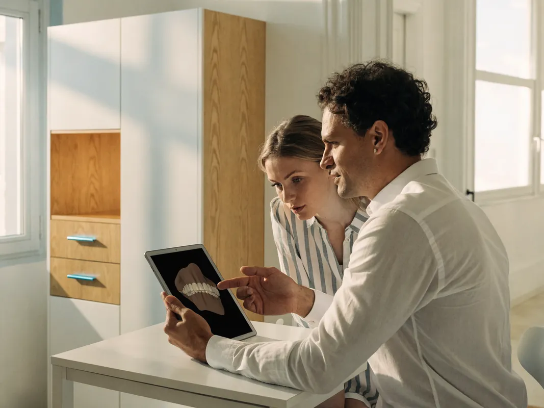
After records are submitted, the actual workflow should identify each responsible party:
- Clinical record review — the provider should name the registered clinician who reviews the records and state the limits of a remote opinion.
- Written treatment plan — sent back to you in your preferred language, including:
- Recommended treatment types
- Brand options (with pros/cons for your case)
- Timing across one or multiple trips
- Hotel and transfer logistics
- Individual quotation
- Specific questions answered
- Follow-up WhatsApp conversation — your questions on the plan, scheduling, modifications.
- Booking — obtain written appointment status plus the named provider, hotel and transfer details.
Do not assume a clinician has reviewed anything until the response names the responsible clinician and distinguishes a provisional discussion from diagnosis.
Common Issues and Solutions
"My dentist won't give me my X-ray."
You have a legal right to your own medical records in most European countries. Request in writing. If still refused, the radiology centre that took the X-ray usually has an independent archive.
"My X-ray is more than a year old."
Send it with the date if you choose. The named clinician can decide whether it is adequate for preliminary discussion or whether current imaging is required.
"My X-ray is on physical film, no digital."
Photograph it on a backlit screen as described above. Or visit a radiology centre to scan the film into digital format.
"I cannot get a panoramic X-ray, only individual periapical X-rays."
Send all of them together with notes. Multiple periapical X-rays of the area you want treated, plus any general overview, can be enough for initial consultation.
"I don't have any X-ray and cannot get one before traveling."
If CBCT may be needed, ask the named provider when and where it would be taken, why it is clinically indicated and whether it is included. A preliminary discussion based on photographs must remain provisional until the responsible clinician completes the necessary assessment.
Privacy and Data
Your X-ray and medical information are confidential. WeCare:
- Does not share your information with third parties
- Stores records on encrypted, EU-GDPR-compliant servers
- Deletes images on request after treatment is complete
WhatsApp message encryption is end-to-end. Your X-ray is not visible to anyone outside the conversation.
If you send an X-ray by WhatsApp, require a response that names the reviewing clinician, distinguishes coordination from diagnosis and sets out the limits of remote planning.