A loose, chipped, uncomfortable or changed implant crown needs diagnosis before a solution is chosen. This guide explains the parts, tests, records and options.
An implant crown can look chipped, feel loose, trap food, change colour, become uncomfortable or simply stop matching nearby teeth. Those observations matter, but none identifies the cause by itself. A symptom is a reason for assessment, not a diagnosis and not an automatic instruction to replace the crown. The cause may be in the visible crown, its retention, the abutment, the prosthetic screw, the implant body, the surrounding tissues, the bite or an unrelated nearby structure. The appropriate response can range from monitoring or polishing to repair, reseating, component treatment, remaking the crown or addressing a wider biological problem.
This is a general planning guide, not personal dental advice. It does not determine whether a reader needs treatment or whether travel is appropriate. A named dentist or suitably qualified restorative clinician needs the relevant history, examination and records. Urgent symptoms need prompt local care rather than an online replacement discussion.
Assessment Before Replacement
Start with the question “what has changed and why?” rather than “how quickly can this crown be remade?” A restoration can be replaced without correcting the reason it changed. If the real problem is screw loosening, an ill-fitting component, occlusal overload, food impaction, inflammation or loss of supporting tissue, a visually new crown may not resolve it. Diagnosis-first planning protects both the implant system and the patient from an irreversible decision based on appearance alone.
The history should establish when the change was first noticed, whether it is stable or progressing, whether it follows trauma or dental work, whether chewing or speech is affected, and whether there is pain, swelling, bleeding, discharge, unpleasant taste, mobility, altered sensation or a change in the bite. Medical history, medicines, smoking, oral-hygiene practices, clenching or grinding, previous periodontal disease and past implant maintenance can matter. This information does not diagnose the cause; it helps the clinician select an examination and decide whether imaging or another test is justified.
Ask the clinician to state a working diagnosis, the evidence supporting it, what remains uncertain and which part is believed to be affected. Ask for alternatives, including no intervention with review when that is reasonable. If the crown has to be removed for diagnosis, ask about the possibility of damage during removal, the route if the screw cannot be accessed, and what temporary arrangement is possible. Consent should reflect those uncertainties before an irreversible step begins.
Crown, Abutment, Screw and Implant Are Different
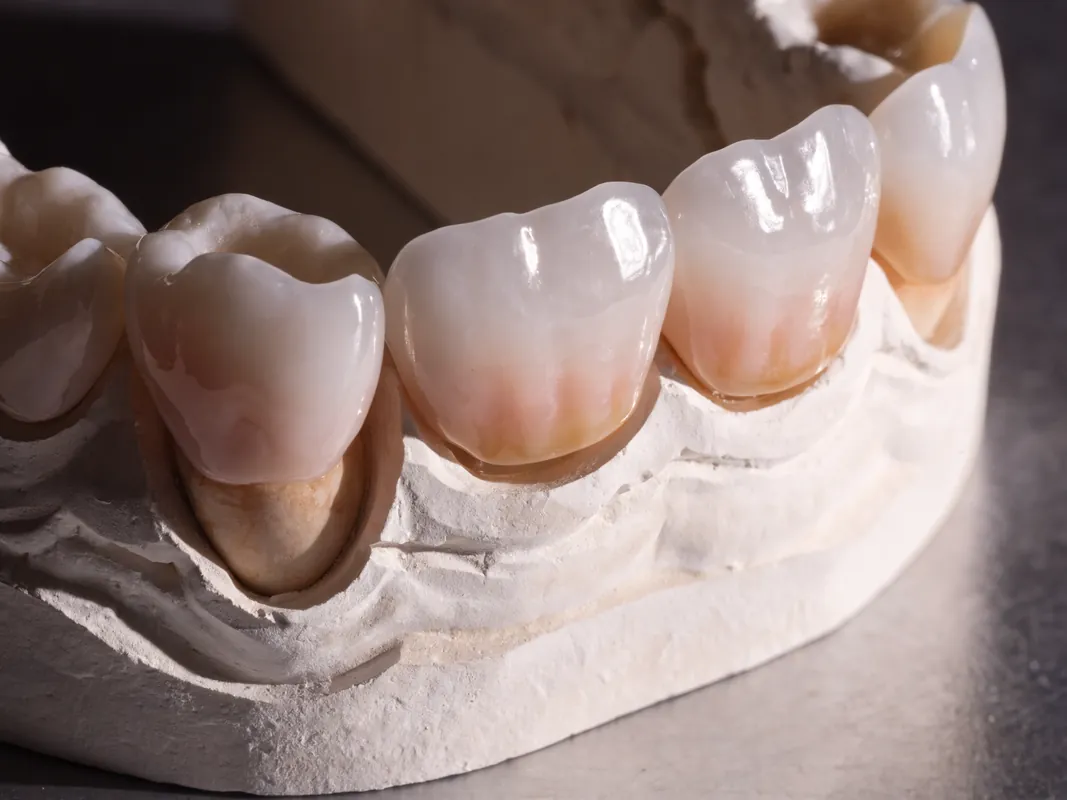
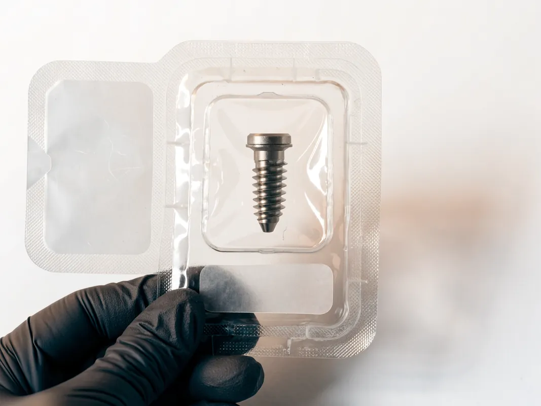
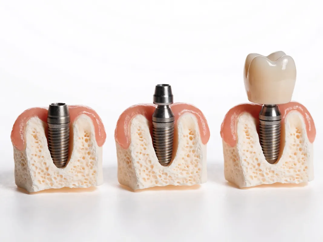
An implant crown, abutment, prosthetic screw and implant body are different parts. The crown is the visible restoration that forms the tooth shape. An abutment can connect that restoration to the implant body. A prosthetic screw may hold an abutment or screw-retained restoration. The implant body is the device placed in bone. Some designs combine or configure these elements differently, so the original component record matters.
A crown can be cement-retained or screw-retained. In a cement-retained design, loss of retention may allow the crown to loosen or detach while the abutment remains connected. In a screw-retained design, movement may relate to the screw, the restoration, an interface or, less commonly, another component. Movement perceived by the patient cannot safely identify which layer is moving. Repeatedly wiggling the crown or biting on it to test movement can add load and should not replace professional assessment.
The implant body should never be assumed healthy merely because a crown issue appears mechanical. Conversely, a chipped crown does not prove the implant body is damaged. The clinician should localise movement, inspect the restoration and tissues, assess the connection and compare with previous records. The FDA explains the general distinction between implant body, abutment and fixation screw at https://www.fda.gov/medical-devices/dental-devices/dental-implants-what-you-should-know. That United States patient source is useful for basic anatomy, not for diagnosing an individual case or establishing device status elsewhere.
Symptoms That Need Review, Not Automatic Replacement
A change in bite, clicking, rocking, roughness, a visible chip, a gap, food packing, bleeding during cleaning, altered contour, persistent bad taste, colour mismatch, recession, a dark area, difficulty flossing, discomfort on chewing or a detached crown all justify a dental review. The urgency depends on the complete presentation. A small surface roughness with no discomfort is different from spreading swelling or difficulty swallowing, but both deserve appropriate professional attention.
Pain around an implant-supported crown can arise from surrounding tissue, pressure, bite, a loose component, fracture, infection, an adjacent tooth, muscle or joint function, or another source. Implants do not have the same pulpal sensation as natural teeth, yet surrounding structures can be painful. A statement such as “implants cannot hurt” is therefore not a safe diagnostic shortcut.
Bleeding can reflect local trauma, plaque-associated inflammation or peri-implant disease, among other possibilities. It should not be covered by a cosmetic remake without tissue assessment. Food trapping can reflect contact, contour, tissue change, neighbouring-tooth movement or cleaning access. A colour concern can result from ceramic, cement, metal display, soft-tissue change, lighting or neighbouring teeth. Each observation is a starting point, not a replacement criterion.
If the crown detaches, retain any recovered part for the dentist and do not use household adhesive. Avoid assuming that recementation is appropriate until the component, fit, retention and underlying tissues are checked. If a fragment may have been inhaled or swallowed and there are respiratory symptoms, seek urgent medical assessment.
Urgent Red Flags
Urgent local assessment is more important than organising elective crown work when there is rapidly increasing swelling, fever with dental symptoms, pus or a persistent unpleasant taste with pain, difficulty opening the mouth, uncontrolled bleeding, significant trauma, spreading facial or neck swelling, or a sudden change in sensation. Difficulty breathing, speaking or swallowing, eye swelling or visual disturbance, or extensive mouth swelling requires emergency help under current NHS advice.
The NHS dental-abscess page at https://www.nhs.uk/conditions/dental-abscess/ explains that a dental abscess needs urgent dental treatment and lists emergency symptoms. It is a general source, not a diagnosis of an implant problem. Follow the current emergency route for the place where the patient is located. Do not delay urgent care while waiting for an overseas provider to answer or while comparing replacement quotations.
A visibly mobile implant body, significant injury or acute bite disruption also needs prompt assessment. The patient may be unable to distinguish a moving crown from a moving implant body, which is precisely why local examination matters. Emergency care may focus on stabilisation and diagnosis rather than definitive cosmetic replacement.
Clinical Examination
The clinician begins by listening to the history and identifying the exact restoration and implant site. Examination may include the crown surface, margins, screw-access area, contacts, contour, occlusion, movement, percussion findings, hygiene access and the surrounding mucosa. The professional may assess plaque, bleeding, suppuration, probing findings and tissue levels using an appropriate technique and compare them with previous records. Adjacent teeth, opposing teeth and other possible sources should also be assessed.
Mobility testing must localise the moving part. A crown, abutment or screw can move even when the implant body is stable. Conversely, apparent stability at one moment does not rule out every component problem. The clinician may need to remove a restoration or access a screw to inspect interfaces. That step requires consent because retrieval can damage ceramic, composite, a screw-access restoration or an underlying component.
Occlusal assessment should consider static contacts, contacts during movement, wear patterns and possible parafunction. A photograph of coloured marking paper is not a complete bite diagnosis. The clinician should relate findings to restoration design, component condition and patient symptoms. If a night appliance exists, its fit and wear may provide context, but it does not prove the cause.
Periodontal and peri-implant assessment is distinct from crown inspection. The EFP guideline at https://www.efp.org/education/continuing-education/clinical-guidelines/guideline-on-treatment-of-peri-implant-diseases/ describes an evidence-based approach to prevention and treatment of peri-implant diseases. The patient information page at https://www.efp.org/for-patients/dental-implants/peri-implant-diseases/ explains peri-implant conditions in accessible language. These sources support professional assessment; they do not authorise self-diagnosis from bleeding or a photograph.
Radiographic Assessment Boundary
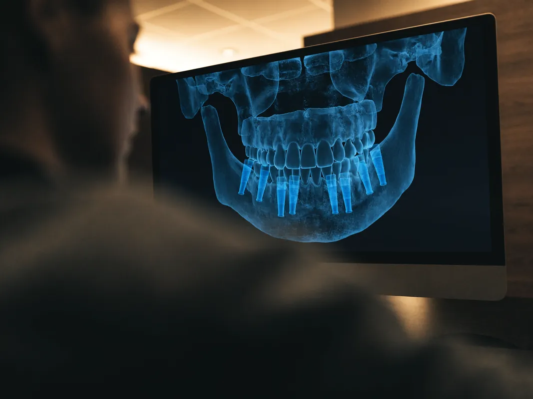
Radiographs can support diagnosis but do not replace clinical examination or identify every mechanical problem. A clinician selects the image type and timing based on the question, previous images, symptoms and radiation justification. Images may help assess bone levels, implant position, certain component relationships, neighbouring structures and some signs of pathology. They may not reveal a small ceramic crack, early screw damage, subtle interface movement, an occlusal cause or every soft-tissue condition.
Comparison with a suitable baseline can be more informative than viewing one isolated image. Ask for the date and reason for the image, what finding it supports, what it cannot show and whether another test would change management. A remote screenshot of a radiograph may lose scale, quality or metadata and should not be presented as a definitive diagnosis.
Three-dimensional imaging is not automatically required for a crown concern. The clinician should justify additional exposure and explain what question the image is intended to answer. Likewise, the absence of obvious radiographic change does not prove that the crown, screw, abutment or bite is healthy. Clinical and radiographic information must be interpreted together.
Screw Loosening and Loss of Retention
Screw loosening can present as movement, clicking, a change in bite, discomfort, a fractured screw-access filling or an intermittent sensation that the tooth is different. Loss of cement retention can produce similar movement in a cement-retained crown. The patient cannot reliably distinguish them. The restoration should be assessed before repeated loading damages a component or surrounding tissue.
The clinician needs the implant and component identity, connection, screw reference, restoration design and relevant manufacturer instructions. Simply tightening a screw without diagnosing why it loosened can miss misfit, wear, damage, occlusal load or an incorrect component. Professional handling should follow the current instruction for use for the exact system and component. A screw that has been damaged, contaminated, repeatedly handled or used outside instructions may require a different decision, which belongs to the responsible clinician.
For a cement-retained crown, reseating depends on fit, abutment condition, retention design, tissue health and the ability to manage excess cement. A detached crown should be inspected for fracture or distortion. Recementing a crown that no longer fits or that masks tissue disease is not a neutral repair.
The ITI consensus source on mechanical and technical risks at https://academy.iti.org/academy/consensus-database/consensus-statement/-/consensus/mechanical-and-technical-risks/1203 supports regular maintenance and attention to technical risk. It is a professional consensus resource, not a fixed rule for an individual crown.
Screw Fracture and Component Damage
A fractured prosthetic screw can leave a fragment inside an implant or abutment. The restoration may be loose, displaced or unexpectedly stable depending on the fracture and design. Retrieval requires appropriate instruments, magnification and judgement. Attempts without knowing the system can damage internal threads or the connection. The clinician should identify the component, inspect interfaces and explain retrieval risks and alternatives.
If internal threads or the connection are damaged, the decision can extend beyond the crown. Options may involve specialist retrieval, a different restorative design, use of a manufacturer-supported component pathway, or wider treatment planning when a component cannot be restored safely. The patient should receive a written explanation of what is known, which parts are reusable, and which records or photographs document the damage.
Component fracture also raises vigilance questions. Retain the reference, lot, removed part and photographs when appropriate. Ask whether the manufacturer should be notified and whether a field safety notice or market action applies to that exact item. The MHRA collection at https://www.gov.uk/government/collections/medical-devices-guidance-for-manufacturers-on-vigilance explains post-market surveillance and field safety activity in Great Britain. Its jurisdiction does not replace the reporting route where the original care occurred.
Chipping, Fracture and Material Loss
A small rough area, superficial chip, loss of veneering material, crack or major fracture can have different implications. The clinician should identify the restorative material and design, determine whether a fracture is limited to a surface layer or involves the supporting structure, inspect for sharp edges, evaluate occlusion and check whether the restoration remains cleanable and structurally serviceable.
Minor material loss may sometimes be polished or repaired, but the feasibility depends on location, material, thickness, appearance, load and remaining support. A repair can be a definitive choice, an interim measure or inappropriate. The expected limitations and maintenance should be explained. A fracture near a screw access or margin may affect retrieval and sealing differently from a small chip away from a functional contact.
Replacing the crown without investigating why it fractured can repeat the same mechanical conditions. The laboratory and clinician should review design, material support, thickness, component fit, screw channel, contact and occlusion. The ITI consensus source at https://academy.iti.org/academy/consensus-database/consensus-statement/-/consensus/survival-and-complication-rates-of-implant-supported-fixed-prostheses-over-the-last-decades/1317 discusses technical complications, tracking and maintenance. It should be read as evidence context, not a promise about one restoration.
Wear, Roughness and Surface Change
Implant crowns and opposing teeth can change through wear, polishing, adjustment, material loss or deposits. A rough surface may retain plaque, irritate the tongue or affect opposing enamel, but the correct response depends on material and cause. Professional polishing may be possible for some surfaces. An aggressive adjustment can alter contour, contact or material thickness, so it should be planned and documented.
Wear patterns can indicate parafunction, an unstable occlusal relationship, material interaction or ordinary individual variation. The clinician should assess the whole bite rather than viewing one crown in isolation. If protective appliance therapy is considered, it needs a current diagnosis, appropriate fit and review; it is not proof that the crown no longer needs assessment.
Colour or gloss change alone may be an aesthetic issue rather than structural damage. Surface staining, deposits and changes in nearby teeth should be distinguished from material alteration. A conservative option may be cleaning or polishing, while a patient may also choose no intervention after understanding the finding.
Contact, Food Trapping and Contour
Food trapping around an implant crown may relate to an open contact, crown contour, papilla or tissue change, movement of a neighbouring tooth, cleaning access or a wider prosthetic design. It can contribute to discomfort and plaque accumulation. It does not tell the patient whether the crown should be repaired or remade.
Assessment should record where food enters, whether the contact is open, whether floss passage has changed, the contour and emergence profile, tissue findings and the position of neighbouring teeth. A contact can sometimes be modified or repaired, while other situations need a new restoration or treatment of a neighbouring tooth. Tissue loss may not be restored simply by making a larger crown.
The patient should receive practical hygiene advice tailored to the restoration. An over-contoured remake can worsen cleaning even if it closes a visible space. Aesthetic pressure should not override tissue access or component constraints. Ask the clinician and laboratory to explain the design objective and how the new or repaired contour will be reviewed.
Occlusion, Bite and Parafunction
A crown that feels “high,” contacts earlier than before, causes a slide, or becomes uncomfortable under load needs occlusal assessment. The cause may be restoration movement, screw loosening, material change, neighbouring-tooth movement, muscle or joint factors, or another dental condition. A bite adjustment made without identifying movement can remove material while leaving the real problem untreated.
The clinician should evaluate the implant crown in the context of the full dentition, functional movements, opposing material and any clenching or grinding history. Adjustment, polishing, repair or remake may be considered depending on the diagnosis. Repeated adjustments that do not resolve symptoms should prompt reassessment rather than an assumption that more material removal is the answer.
Parafunction can affect technical risk, but patients should not be blamed for a complication. The plan should discuss modifiable factors, restoration design, maintenance and realistic uncertainty. An appliance may be one part of management where appropriate; its indication and review are clinical decisions.
Aesthetic Concerns and Expectations
A crown may no longer match because neighbouring teeth change, tissue recedes, lighting reveals opacity, metal or an abutment becomes visible, the crown shape appears different, or the patient’s goals change. Aesthetic dissatisfaction deserves a respectful assessment, but elective replacement is not the only response. Cleaning, polishing, tissue evaluation, photography under consistent conditions, limited repair, accepting the appearance or wider restorative planning may be discussed.
The clinician should explain what a new crown can and cannot change. Replacing ceramic cannot reliably recreate lost tissue, move an implant body or assure symmetry. Altering one crown may reveal differences in adjacent teeth. A trial design, shade record or laboratory consultation can support consent without promising an exact appearance.
If the existing crown is biologically and mechanically serviceable, replacement for appearance is an elective decision with its own removal risks. Ask how the crown will be retrieved, whether the abutment can be preserved, what happens if a component is damaged and what temporary solution is possible. The choice should not be rushed by travel arrangements.
Soft Tissue and Peri-Implant Disease
Redness, swelling, bleeding, suppuration, recession, tenderness or changing tissue contours require periodontal and peri-implant assessment. Peri-implant mucositis and peri-implantitis are clinical diagnoses, not labels a patient should apply from one symptom. The EFP guideline emphasises diagnosis, risk-factor control, professional intervention and supportive care. Treatment depends on disease findings, not simply on replacing the visible crown.
Crown contour, margin position, cement remnants and hygiene access can be relevant to tissue health, but they are not the only causes. A clinician may need to remove the restoration to improve access or investigate a factor. That does not mean a new crown alone treats the disease. The plan should separate disease management from restorative modification and explain how the two interact.
Baseline records help interpret change. Ask for prior radiographs, probing information, photographs and maintenance notes when available. A local dentist should not be expected to accept a remote statement that tissues are healthy without performing an independent assessment.
Repair, Polish, Monitor or Reseat
Conservative management may be reasonable when the diagnosis and material allow it. Monitoring can be appropriate for a stable, harmless finding if the patient understands what change should trigger review. Polishing can address certain rough surfaces. Intraoral or laboratory repair may restore limited material loss. Reseating may be considered after loss of retention when the restoration, component and tissues are suitable.
Each option needs a defined aim, limitation and review plan. “Repairable” is not a universal property of chip size. Access, load, appearance, material, bonding substrate, contamination and support matter. A repair may change surface quality or colour. Reseating may not correct the original retention problem. Monitoring should not become neglect when disease or progressive damage is suspected.
Ask what evidence supports the selected option and what would cause the plan to change. Obtain a record of the material and component used. If a screw is handled, the clinician should follow current manufacturer instructions for the exact component rather than apply a generic torque assumption.
When Replacement May Be Considered
Replacement may be considered when the crown is structurally unserviceable, cannot be repaired to an acceptable form, repeatedly loses retention after the cause has been assessed, cannot provide suitable contact or contour, prevents necessary access, has an irretrievable aesthetic limitation that the patient chooses to address, or forms part of a wider diagnosed restorative problem. These are decision categories, not rules.
Before replacement, the clinician should identify whether the existing restoration is screw-retained or cement-retained, how it can be removed, which underlying parts may be preserved, and whether the component system remains obtainable. The implant body and surrounding tissues need independent assessment. The laboratory needs an accurate prescription and compatible components.
The written plan should state whether the new item is a crown only, crown plus abutment, a screw-retained unit, a provisional restoration or another configuration. It should identify uncertain findings that could expand the scope after removal. Consent should include the possibility that the crown cannot be removed intact or that underlying damage changes the available options.
Non-Restorable Findings and Wider Alternatives
Sometimes assessment identifies damage or biological disease that cannot be solved with a new crown. Internal connection damage, an unrestorable implant position, severe component fracture, loss of integration, progressive tissue disease, inadequate restorative space or a broader prosthetic problem may require specialist planning. The appropriate alternative depends on the diagnosis and patient priorities.
Options can include specialist component retrieval, a modified abutment or restorative pathway supported by current instructions, a removable restoration, a tooth-supported option where appropriate, leaving a space, managing disease before restoration, or removal of an implant body when clinically indicated. This is not a menu for self-selection. Each option has different risks, maintenance and reversibility.
Ask the clinician to distinguish salvage of the crown, salvage of the abutment or connection, and management of the implant body. Seek another opinion when the diagnosis is unclear or the proposed intervention is irreversible. A known manufacturer name does not eliminate the need for this analysis.
Component Record, Implant Passport and Serviceability
Future care depends on identifying the implant system and restorative components. Request the legal manufacturer, implant family, connection, implant reference and lot, abutment and screw references, restoration design, placement site and date, and any component changed during maintenance. An implant passport, product-label copy and operative record can make this information portable.
The record should distinguish the implant body from the abutment, screw and crown. A broad phrase such as “titanium implant with zirconia crown” is not enough for retrieving a fractured screw or selecting a compatible scan body. Keep the original label and a clear English summary if treatment records use another language.
Serviceability also depends on component availability, tools, professional willingness and the condition of the device. A local dentist may need to contact the manufacturer or refer to a clinician familiar with the system. No patient should be told that every dentist can service every implant. Check this before elective travel and before an old crown is removed.

Laboratory and Material Records
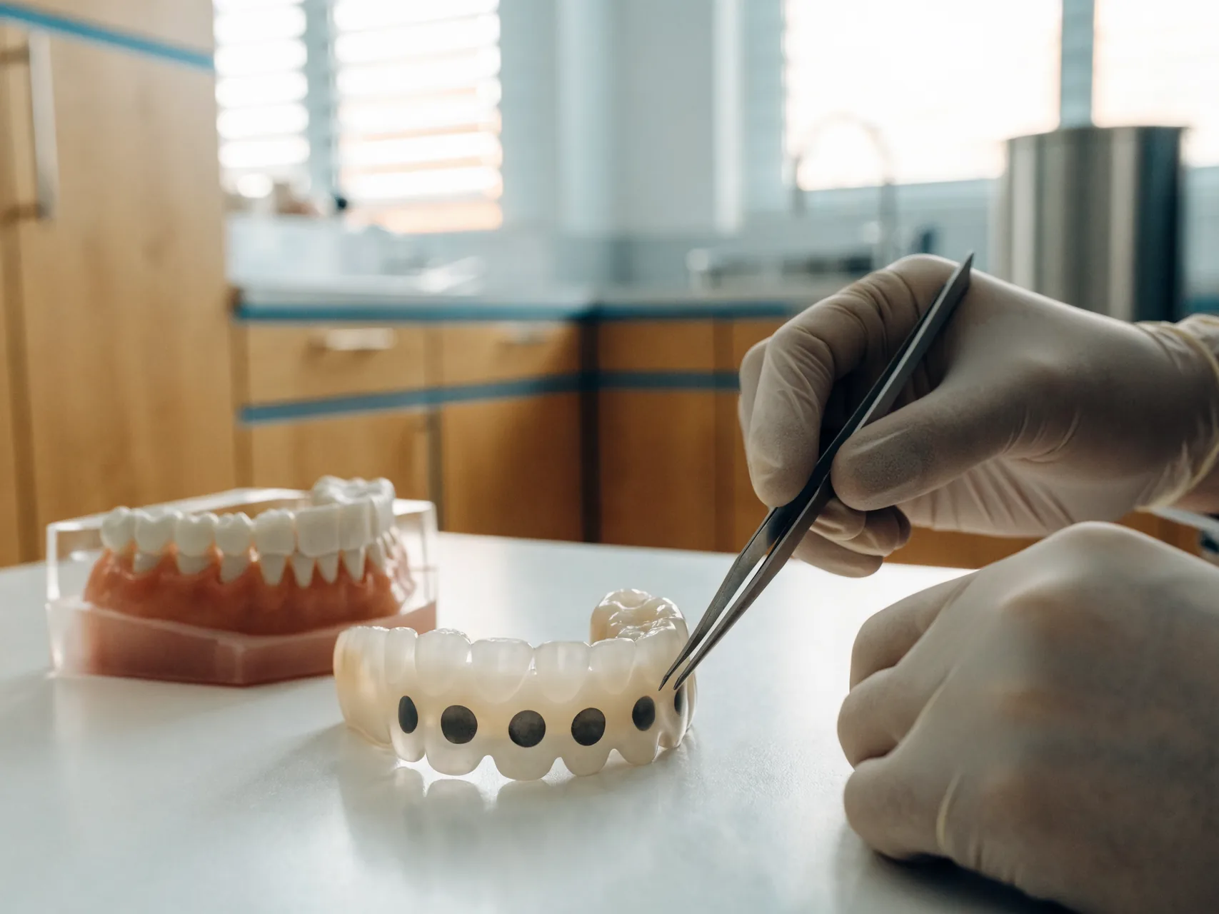
The laboratory prescription should identify the restoration material, framework or monolithic design, abutment or titanium base, connection, screw channel, shade, contact and occlusal instructions, and responsible clinician. Ask whether the laboratory is remaking an existing design or creating a new one. Digital design files can help but must be matched to the correct component library and physical parts.
For a fracture or chip, ask the laboratory to document the material layers and observed failure. For a detached crown, record whether cement remained on the crown or abutment and whether fit was verified. For a remake, ask which components are reused and which are new. The invoice should not be the only technical record.
A material label does not determine clinical suitability or resolve a mechanical cause. The clinician and dental technician have separate professional roles and should communicate through a clear prescription. Patients should know who is responsible for clinical decisions and who manufactures the restoration.
Consent and Itemised Quotation
A written proposal should begin with the diagnosis and parts affected. It should list examination or imaging already completed, uncertainty that remains, proposed removal method, repair or replacement scope, component and laboratory details, alternatives, follow-up, exclusions, complaint route and financial terms. If travel is involved, separate clinical responsibilities from non-clinical arrangements.
The quotation should identify whether it covers assessment only, repair, crown removal, a new crown, abutment, screw, provisional restoration, laboratory work, imaging, tissue treatment or follow-up. Avoid a single line called “implant crown replacement” when several parts may be needed. Ask how a change discovered after crown removal is approved and priced before additional work proceeds.
Consent is an ongoing conversation. The patient should understand that a remote proposal is provisional, that removal can reveal new findings, and that deciding not to proceed remains possible. A deposit or booked journey should not become a clinical deadline.
Maintenance and Local Dental Handover
Long-term care needs a named local route. Before treatment abroad, ask a UK dentist or relevant professional whether they can assess the existing implant, what records they require and whether referral is more appropriate. They remain responsible for their own diagnosis and are not obliged to adopt an overseas plan.
Maintenance can include professional assessment of tissues, plaque control, hygiene access, probing where appropriate, restoration condition, contacts, occlusion and patient symptoms. The interval is individualised. Avoid fixed lifespan charts and universal recall schedules because medical, biological, mechanical and behavioural factors differ.
After any repair or replacement, request updated component and laboratory records. Tell the local dentist what changed. If a concern recurs, return to diagnosis rather than assuming the same cause. Current manufacturer notices and regulator information should be checked against exact identifiers when a device issue is suspected.
Travel and No-Travel Decision
Do not book travel for a planned crown replacement before a named clinician has reviewed the available records and explained what remains provisional. A local assessment may show that the crown can be monitored or repaired, that another component is involved, that tissue treatment comes first, or that urgent care is needed. Travel should not delay diagnosis.
If overseas care is still considered, obtain legal provider and clinician identities, component availability, a written plan, removal contingencies, laboratory scope, aftercare, record handover and complaint process. The GDC guide at https://www.gdc-uk.org/standards-guidance/information-for-patients-public/going-abroad-for-dental-treatment explains questions UK patients should ask and the limits of UK regulatory reach. Recheck the guidance before committing.
Do not travel when acute symptoms need local urgent care, when the diagnosis is not established, when necessary records are unavailable, when a component cannot be sourced, when a medical professional advises against travel, or when the plan relies on a fixed outcome from an unseen examination. A person fit to travel still needs an appropriate treatment plan; those are separate decisions.
Diagnostic and Decision Workbook
Use the following prompts with the clinician. The repeated structure is intentional: it keeps the observation, evidence and decision boundary separate so that a sales summary cannot quietly become a diagnosis.
Primary concern
Assessment question: What exactly changed from the patient’s baseline and when was it first noticed?
Evidence to request: A dated symptom history, photographs where useful and previous notes for comparison.
Decision boundary: The observation defines the assessment question but does not identify the affected component.
Ask the clinician to connect the answer to the named component and examination finding. Retain it with the dated plan so another professional can understand the reasoning without relying on an informal message.
Urgency screen
Assessment question: Are there swelling, fever, discharge, bleeding, trauma, breathing or swallowing concerns that change urgency?
Evidence to request: A contemporaneous clinical triage record and the advice given for local care.
Decision boundary: Urgent risk is addressed before elective restoration planning or travel.
If the answer changes after examination or crown removal, request the reason, revised scope and alternatives before further irreversible work. Travel or prior payment should not remove that decision point.
Restoration identity
Assessment question: Is the visible unit a single crown, part of a bridge or part of another implant-supported reconstruction?
Evidence to request: The original treatment plan, laboratory prescription and current clinical chart.
Decision boundary: The scope determines which adjacent units and components may need assessment.
A vague answer is an unresolved dependency. Do not fill the gap with a lifespan chart, brand reputation or assumption that a known component can be serviced anywhere.
Retention design
Assessment question: Is the restoration screw-retained, cement-retained or configured through another component pathway?
Evidence to request: A clinical record, radiograph where justified, screw-access information and laboratory note.
Decision boundary: Removal and reseating risks differ by retention design and cannot be inferred from appearance.
Record both what is known and what the available evidence cannot establish. That distinction reduces unnecessary crown replacement and helps a later local professional continue care safely.
Movement localisation
Assessment question: Which part moves during professional examination: crown, abutment, screw interface or implant body?
Evidence to request: A signed examination finding and any supporting image or component inspection.
Decision boundary: The solution depends on localising movement rather than treating every loose sensation as a crown problem.
The responsible professional should sign or otherwise authenticate the conclusion. A photograph or radiograph can support it, but neither becomes a diagnosis without clinical interpretation.
Implant system identity
Assessment question: What legal manufacturer, implant family and connection are present?
Evidence to request: Implant passport, label copy, operative record and manufacturer-confirmed reference where needed.
Decision boundary: Exact identity supports compatible instruments, screws, scan bodies and abutments.
Ask the clinician to connect the answer to the named component and examination finding. Retain it with the dated plan so another professional can understand the reasoning without relying on an informal message.
Crown material
Assessment question: What crown and framework materials were used, and is the design monolithic, layered or otherwise constructed?
Evidence to request: Laboratory prescription, material label, invoice detail and design record.
Decision boundary: Repairability and polishing methods depend on material and where damage occurred.
If the answer changes after examination or crown removal, request the reason, revised scope and alternatives before further irreversible work. Travel or prior payment should not remove that decision point.
Abutment identity
Assessment question: Is the abutment stock, custom or integrated into another restorative component?
Evidence to request: Abutment manufacturer, reference, material and laboratory or clinical record.
Decision boundary: A crown remake may not solve damage or misfit at the abutment level.
A vague answer is an unresolved dependency. Do not fill the gap with a lifespan chart, brand reputation or assumption that a known component can be serviced anywhere.
Screw identity
Assessment question: Which prosthetic screw is present and what current manufacturer instructions apply?
Evidence to request: Exact screw reference, connection and applicable instruction for use.
Decision boundary: Professional retrieval or handling depends on the actual component rather than a generic torque value.
Record both what is known and what the available evidence cannot establish. That distinction reduces unnecessary crown replacement and helps a later local professional continue care safely.
Screw-access condition
Assessment question: Is the access restoration intact, fractured, stained or hiding evidence of movement?
Evidence to request: Clinical photographs and examination of the access area before intervention.
Decision boundary: Access condition can suggest a technical concern but does not diagnose its cause.
The responsible professional should sign or otherwise authenticate the conclusion. A photograph or radiograph can support it, but neither becomes a diagnosis without clinical interpretation.
Cement-retention condition
Assessment question: If cement-retained, is there loss of retention, fit change or suspected residual cement?
Evidence to request: Examination, tissue assessment and inspection after removal if clinically justified.
Decision boundary: Recementation is considered only after fit, abutment and tissue conditions are understood.
Ask the clinician to connect the answer to the named component and examination finding. Retain it with the dated plan so another professional can understand the reasoning without relying on an informal message.
Chip classification
Assessment question: Is material loss superficial, within a veneering layer or through the supporting structure?
Evidence to request: Magnified clinical assessment, photographs and laboratory material information.
Decision boundary: Depth and location influence whether polishing, repair or remake is technically reasonable.
If the answer changes after examination or crown removal, request the reason, revised scope and alternatives before further irreversible work. Travel or prior payment should not remove that decision point.
Crack assessment
Assessment question: Is a visible line a superficial mark, a crack or a feature of the material and lighting?
Evidence to request: Professional inspection, transillumination or other justified tests and laboratory context.
Decision boundary: A photograph alone can overstate or miss structural damage.
A vague answer is an unresolved dependency. Do not fill the gap with a lifespan chart, brand reputation or assumption that a known component can be serviced anywhere.
Surface roughness
Assessment question: What caused the rough area and can it be polished without compromising contour or material?
Evidence to request: Material identification, surface examination and record of any prior adjustment.
Decision boundary: Conservative finishing may help some surfaces while another defect needs a different plan.
Record both what is known and what the available evidence cannot establish. That distinction reduces unnecessary crown replacement and helps a later local professional continue care safely.
Wear pattern
Assessment question: Is wear isolated to the implant crown or present across opposing and neighbouring teeth?
Evidence to request: Occlusal examination, photographs, appliance history and previous records.
Decision boundary: A system-wide pattern should not be treated as an isolated cosmetic defect.
The responsible professional should sign or otherwise authenticate the conclusion. A photograph or radiograph can support it, but neither becomes a diagnosis without clinical interpretation.
Static bite contacts
Assessment question: Does the implant crown contact prematurely or differently in the current bite?
Evidence to request: Documented occlusal assessment rather than a patient sensation alone.
Decision boundary: Adjustment should follow localisation of movement and the diagnosis.
Ask the clinician to connect the answer to the named component and examination finding. Retain it with the dated plan so another professional can understand the reasoning without relying on an informal message.
Excursive contacts
Assessment question: What happens during lateral and forward movements, and are symptoms reproduced?
Evidence to request: Professional functional examination and recorded findings.
Decision boundary: Dynamic loading can influence the technical plan but is not measured by one static mark.
If the answer changes after examination or crown removal, request the reason, revised scope and alternatives before further irreversible work. Travel or prior payment should not remove that decision point.
Parafunction context
Assessment question: Are there reported or observed signs of clenching or grinding that affect risk management?
Evidence to request: History, wear signs and appliance examination where relevant.
Decision boundary: Parafunction is one factor and does not transfer responsibility away from proper design and follow-up.
A vague answer is an unresolved dependency. Do not fill the gap with a lifespan chart, brand reputation or assumption that a known component can be serviced anywhere.
Interproximal contact
Assessment question: Is an open or weak contact contributing to food impaction or tissue irritation?
Evidence to request: Contact assessment, neighbouring-tooth position and patient history.
Decision boundary: Closing a contact may require repair, remake or treatment of another tooth depending on cause.
Record both what is known and what the available evidence cannot establish. That distinction reduces unnecessary crown replacement and helps a later local professional continue care safely.
Emergence contour
Assessment question: Can the patient clean the crown contour and does it support healthy tissue access?
Evidence to request: Clinical inspection, hygiene assessment and photographs or scans where useful.
Decision boundary: Increasing contour for appearance can worsen access and should be justified.
The responsible professional should sign or otherwise authenticate the conclusion. A photograph or radiograph can support it, but neither becomes a diagnosis without clinical interpretation.
Tissue inflammation
Assessment question: Are redness, bleeding or swelling associated with plaque, contour, cement, disease or another factor?
Evidence to request: Periodontal and peri-implant findings interpreted by a qualified clinician.
Decision boundary: A restorative change is only one possible part of disease management.
Ask the clinician to connect the answer to the named component and examination finding. Retain it with the dated plan so another professional can understand the reasoning without relying on an informal message.
Suppuration or discharge
Assessment question: Is there clinical evidence of suppuration, a sinus tract or another source of discharge?
Evidence to request: Urgent clinical assessment, appropriate imaging and diagnosis.
Decision boundary: This finding changes urgency and should not be managed as an elective aesthetic remake.
If the answer changes after examination or crown removal, request the reason, revised scope and alternatives before further irreversible work. Travel or prior payment should not remove that decision point.
Probing and baseline
Assessment question: How do current peri-implant findings compare with valid previous measurements?
Evidence to request: Baseline charting, current professional measurements and context for technique.
Decision boundary: Change over time can be more informative than an isolated value.
A vague answer is an unresolved dependency. Do not fill the gap with a lifespan chart, brand reputation or assumption that a known component can be serviced anywhere.
Radiographic question
Assessment question: What specific diagnostic question is an image intended to answer?
Evidence to request: The image, report, date, indication and comparison with a suitable baseline.
Decision boundary: Imaging is justified by a question and cannot reveal every technical or soft-tissue problem.
Record both what is known and what the available evidence cannot establish. That distinction reduces unnecessary crown replacement and helps a later local professional continue care safely.
Adjacent-tooth assessment
Assessment question: Could pain, food trapping or bite change arise from a neighbouring natural tooth?
Evidence to request: Examination and diagnostic tests of adjacent structures.
Decision boundary: Avoiding tunnel vision prevents unnecessary intervention on an intact implant crown.
The responsible professional should sign or otherwise authenticate the conclusion. A photograph or radiograph can support it, but neither becomes a diagnosis without clinical interpretation.
Aesthetic objective
Assessment question: Which feature concerns the patient: shade, opacity, contour, tissue display, symmetry or surface?
Evidence to request: Standardised photographs, patient discussion and laboratory communication.
Decision boundary: A specific objective allows honest discussion of what a new crown cannot change.
Ask the clinician to connect the answer to the named component and examination finding. Retain it with the dated plan so another professional can understand the reasoning without relying on an informal message.
Removal pathway
Assessment question: How can the existing crown be removed and what damage is possible during retrieval?
Evidence to request: Retention design, access plan, instruments and written contingency.
Decision boundary: Removal is not risk-free and can expose underlying conditions that change scope.
If the answer changes after examination or crown removal, request the reason, revised scope and alternatives before further irreversible work. Travel or prior payment should not remove that decision point.
Repair feasibility
Assessment question: Can the defect be polished or repaired while preserving strength, cleanability and appearance?
Evidence to request: Material-specific assessment and a written explanation of limitations.
Decision boundary: A repair decision considers function and maintenance, not only whether material can be added.
A vague answer is an unresolved dependency. Do not fill the gap with a lifespan chart, brand reputation or assumption that a known component can be serviced anywhere.
Reseating feasibility
Assessment question: If the crown detached, does it still fit and is the abutment and tissue condition suitable?
Evidence to request: Inspection of crown, abutment, retention surfaces and surrounding tissues.
Decision boundary: A quick reseat can reproduce the original problem when its cause remains unresolved.
Record both what is known and what the available evidence cannot establish. That distinction reduces unnecessary crown replacement and helps a later local professional continue care safely.
Replacement scope
Assessment question: Would the proposed work replace only the crown or also an abutment, screw or other component?
Evidence to request: An itemised component list and site-specific laboratory prescription.
Decision boundary: Precise scope prevents a crown label from concealing wider intervention.
The responsible professional should sign or otherwise authenticate the conclusion. A photograph or radiograph can support it, but neither becomes a diagnosis without clinical interpretation.
Laboratory responsibility
Assessment question: Which laboratory will repair or remake the restoration and what component library will it use?
Evidence to request: Legal laboratory identity, prescription, material record and validated component information.
Decision boundary: A later clinician needs to trace who made the restoration and with which parts.
Ask the clinician to connect the answer to the named component and examination finding. Retain it with the dated plan so another professional can understand the reasoning without relying on an informal message.
Component availability
Assessment question: Can the exact screw, abutment, scan body and instruments be sourced where care will occur?
Evidence to request: Dated manufacturer or supplier confirmation matched to exact references.
Decision boundary: Known corporate branding does not establish access to an older or regional component.
If the answer changes after examination or crown removal, request the reason, revised scope and alternatives before further irreversible work. Travel or prior payment should not remove that decision point.
Field safety check
Assessment question: Does any current official notice apply to the exact reference, lot and market?
Evidence to request: Manufacturer correspondence and regulator search records with dates.
Decision boundary: A notice for another product or market should not be generalised to this case.
A vague answer is an unresolved dependency. Do not fill the gap with a lifespan chart, brand reputation or assumption that a known component can be serviced anywhere.
Local handover
Assessment question: Which local dentist can assess the case and what records do they need?
Evidence to request: A named route, record list and acknowledgement that acceptance follows independent assessment.
Decision boundary: Cross-border serviceability depends on professional willingness as well as component access.
Record both what is known and what the available evidence cannot establish. That distinction reduces unnecessary crown replacement and helps a later local professional continue care safely.
Consent boundary
Assessment question: What remains uncertain until removal or further examination, and how will a change be authorised?
Evidence to request: A written consent process, alternatives and a pause point before expanded work.
Decision boundary: The patient should not discover a materially wider intervention after an irreversible step without discussion.
The responsible professional should sign or otherwise authenticate the conclusion. A photograph or radiograph can support it, but neither becomes a diagnosis without clinical interpretation.
Itemised quotation
Assessment question: Which examination, removal, repair, component, laboratory and follow-up items are included or excluded?
Evidence to request: A dated line-by-line quotation tied to the clinical plan.
Decision boundary: Equivalent scope can be compared without turning price into proof of clinical quality.
Ask the clinician to connect the answer to the named component and examination finding. Retain it with the dated plan so another professional can understand the reasoning without relying on an informal message.
Review plan
Assessment question: What findings will be reviewed after monitoring, repair, reseating or replacement?
Evidence to request: A patient-specific follow-up plan and updated records.
Decision boundary: A defined review closes the diagnostic loop instead of treating intervention as the end of care.
If the answer changes after examination or crown removal, request the reason, revised scope and alternatives before further irreversible work. Travel or prior payment should not remove that decision point.
Frequently Asked Questions
Does every loose implant crown need a new crown?
No. Movement can arise from cement retention, a prosthetic screw, an abutment, the crown structure or the implant body. A clinician must localise the moving part, inspect the system and diagnose the cause before choosing repair, reseating, component care or replacement.
Can I tell whether the implant body is loose at home?
Not reliably. A moving restoration can feel similar to deeper movement, and repeated self-testing may add load. Arrange professional assessment, and seek prompt local care when movement is accompanied by significant pain, swelling, trauma or other urgent symptoms.
Is a chipped ceramic surface always a reason to remake the crown?
No. The material, depth, location, supporting design, roughness, bite and appearance all matter. Some defects may be polished or repaired, while others may make a remake reasonable. The clinician should also investigate why the chip occurred.
What if my implant crown has completely come off?
Keep the recovered restoration for the dentist and avoid household adhesive. The clinician should inspect the crown, abutment, retention, fit, tissues and component identity before deciding whether reseating, repair or a new restoration is appropriate.
Why does the exact implant system matter for crown care?
The implant connection determines compatible screws, abutments, scan bodies, drivers and restorative components. An implant passport, label copy and component schedule reduce guesswork and support safer maintenance or referral when care occurs in another country.
Can an X-ray show whether the crown is cracked?
Not every crack or technical problem is visible on a radiograph. Imaging can answer selected questions about bone and component relationships, but material inspection, movement localisation, tissue assessment and bite examination remain necessary.
Does bleeding around an implant mean the crown should be changed?
No. Bleeding needs periodontal and peri-implant assessment. Plaque-associated inflammation, contour, cement remnants, tissue disease or another factor may be involved. Restorative modification may be relevant, but a new crown alone is not a universal treatment.
Can a dentist simply tighten a loose implant screw?
The dentist first needs to identify the system and diagnose why movement occurred. Component condition, fit, occlusion and current manufacturer instructions matter. Blind tightening can overlook damage, misfit or the wrong component.
What records should I bring to an implant-crown review?
Bring the implant passport or label, operative record, prior radiographs, crown and abutment details, laboratory prescription, maintenance notes and any record of previous screw or crown work. Original documents are preferable to a typed brand name.
Can a small food trap be repaired without replacing the crown?
Sometimes, but the cause determines the option. Contact, contour, tissue change and neighbouring-tooth movement should be assessed. A repair, remake, monitoring or care involving another tooth may be discussed after diagnosis.
What if the crown looks darker at the gum line?
The cause may involve tissue recession, material opacity, abutment display, deposits, lighting or another factor. Clinical examination and consistent photographs help clarify the concern and what a restorative change can realistically alter.
Is an implant crown expected to last a fixed number of years?
No fixed lifespan applies to every restoration. Material, design, component fit, bite, tissues, maintenance, health and events such as trauma vary. Review current condition and risk rather than using a calendar as a replacement instruction.
Should I replace an implant crown before it develops a problem?
Routine elective replacement is not justified by age alone. A clinician should assess function, tissues, material, components, hygiene and patient goals. Monitoring can be reasonable when findings are stable and serviceable.
Can polishing solve every rough or worn implant crown?
No. Polishing depends on the material, damage depth, contour and remaining support. The professional should avoid removing material without understanding the defect and should record any adjustment for future care.
What happens if the crown cannot be removed intact?
The consent and quotation should address that possibility before retrieval. The clinician may need to section or damage the restoration, and underlying findings can change the component or laboratory scope. A temporary route should be discussed.
Can a UK dentist take over an overseas implant crown case?
A UK dentist decides independently after assessment. They may need exact records, obtainable components, suitable tools or a referral route. A familiar implant name does not create an automatic handover obligation.
When should I avoid travelling for a crown remake?
Do not let travel delay urgent local assessment, and avoid committing while diagnosis, component identity, medical fitness, local follow-up or the revised scope remains unresolved. Seek personal advice from appropriate professionals.
What should an itemised crown proposal include?
It should identify the diagnosis, parts affected, examination and imaging, removal method, repair or replacement scope, components, laboratory, alternatives, uncertainties, follow-up, records, exclusions, complaint route and financial terms.
How are a repair and a replacement compared fairly?
Compare their aims, reversibility, material limits, component implications, cleanability, expected maintenance and what happens if the underlying cause persists. Price and travel convenience do not establish clinical appropriateness.
What should happen after a crown is repaired or remade?
Obtain updated component and laboratory records, follow the personalised hygiene and review plan, and tell the local dentist what changed. Recurrent symptoms need reassessment rather than an assumption that the previous cause has returned.
Official Sources and Currency
The EFP guideline and patient information explain peri-implant disease assessment and treatment principles. ITI consensus pages provide professional context on mechanical and technical risk, component tracking, occlusal review and maintenance. FDA patient information distinguishes implant-system parts. NHS guidance defines urgent abscess and emergency symptoms. GDC guidance frames questions and regulatory limits for UK patients considering care abroad. MHRA material explains post-market surveillance and field safety activity in Great Britain.
Each source has a jurisdiction, audience and publication context. Recheck the live page and any linked primary guideline near the date of assessment. Do not convert a consensus statement, patient webpage, device listing or manufacturer instruction into a personal diagnosis. The final decision belongs to appropriately qualified professionals using current records and examination findings.
The practical rule is diagnosis before replacement: identify the affected part, urgent risk, biological condition, mechanical cause, restoration material, component system and local service route; then compare monitoring, repair, reseating, component intervention, replacement and wider alternatives through informed consent.