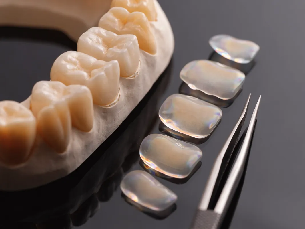
Root canal treatment, also called endodontic treatment, may preserve a tooth when the pulp inside it is inflamed or infected and the remaining tooth can be restored. Treatment generally removes the affected pulp, cleans and disinfects the internal canal space, then fills and seals it. A filling, onlay or crown may be needed afterwards to protect and restore function; the right restoration depends on the tooth and how much sound structure remains.
Symptoms alone do not confirm the diagnosis. Toothache, lingering temperature sensitivity, pain on biting, a darkened tooth, a gum spot or swelling can have several causes. Eligibility for root canal treatment therefore depends on an examination, appropriate tests and a judgement about whether the tooth is restorable.
The [NHS root canal overview](https://www.nhs.uk/tests-and-treatments/root-canal-treatment/) describes removal of inflamed or infected pulp, cleaning and filling of the canal system, and later restoration where needed. The current [NHS England dental care pathways guidance](https://www.england.nhs.uk/long-read/nhs-dentistry-care-pathways-guidance/) places diagnosis, disease control, restorability and the coronal seal inside the same care pathway. The [GDC consent standard](https://standards.gdc-uk.org/pages/principle3/principle3) and [patient-record standard](https://standards.gdc-uk.org/pages/principle4/principle4) are useful question frameworks for UK patients, but they do not certify a clinician or facility in Turkey.
This guide does not diagnose a tooth or prescribe an instrument, irrigant, filling material, antibiotic, pain medicine, restoration, appointment count, price or travel schedule. Those decisions depend on the patient, current findings, the tooth and the named clinician. It is designed to help a patient compare a complete pathway rather than a headline root canal fee.
Start with the decision, not the destination
The real decision is whether the tooth is urgent, whether its pulp or tissues around the root are diseased, whether the tooth can be restored predictably enough to justify treatment, and which reasonable alternatives exist. Travel becomes relevant only after those questions have a provisional answer.
A remote estimate based on one radiograph may omit a crack, decay below a crown, poor gum support, limited remaining tooth tissue or a restoration that must be replaced. Conversely, pain does not automatically mean the pulp must be removed. Ask for the working diagnosis and the evidence supporting it, together with what remains uncertain until direct examination.
Compare the entire tooth-retention pathway: triage, diagnosis, isolation, endodontic procedure, temporary seal if used, definitive restoration, review and contingency care. A cheaper canal procedure can become the more expensive option if the permanent restoration, re-treatment risk or local follow-up is missing from the proposal.
Understand the tooth and the diagnostic question
The visible crown is only part of the tooth. Inside are the pulp chamber and a root canal system that may contain branches, curves, narrow areas and anatomical variation. Disease can involve inflamed living pulp, necrotic tissue, infection within the canal system or tissues around the root. These states are not interchangeable and may lead to different options.
Ask for a plain-language diagnosis that names the tooth and the suspected pulpal and apical condition. The explanation should connect symptoms, clinical tests and images without claiming more certainty than the evidence supports. A response to cold, tapping or biting is one data point, not a treatment plan by itself.
The clinician should also identify the cause where possible: deep decay, a leaking restoration, repeated procedures, trauma, a crack, wear, previous endodontic treatment or another problem. Managing the canal without addressing the cause and the future seal leaves the pathway incomplete.
Symptoms can mislead in both directions
Severe pain may come from pulp inflammation, a crack, bite trauma, gum disease, sinus-related pain or another tooth. A tooth with a dead pulp may produce little pain until infection spreads. A previously treated tooth may be tender because of the bite, restoration, fracture or persistent disease. Self-diagnosis from pain location is unreliable because dental pain can be referred.
Record when symptoms began, whether they are spontaneous or triggered, what makes them better or worse, whether they linger, whether biting or release hurts, and whether swelling, fever, altered taste or a gum spot is present. Mention recent dentistry, injury and pain medicine because each can change the history.
If tests disagree with the symptoms, the answer is not automatically to start treatment. Reassessment, comparison with neighbouring teeth, additional views or referral may be safer than an irreversible procedure on an uncertain tooth.
Urgent symptoms come before travel
An urgent dental problem is usually a poor-value reason to travel. Delay can allow pain or infection to worsen, while flights and unfamiliar follow-up make continuity harder. If there is facial or mouth swelling, fever, difficulty swallowing or breathing, spreading redness, severe worsening pain or a dental injury, seek local urgent care. Do not wait for an overseas estimate or routine appointment.
A dental abscess needs prompt assessment. Depending on the findings, urgent care may involve drainage, endodontic treatment, extraction or another measure. Antibiotics are not a substitute for removing the dental source when operative care is required, and they should be prescribed only after an appropriate clinical decision.
Travel may be considered for a stable, non-urgent case or planned re-treatment, but only after local triage has ruled out a time-sensitive problem. The expected saving on one tooth can disappear once travel, time away and local follow-up are counted.
Local stabilisation may be the correct first step
An urgent visit may aim to diagnose, relieve pressure, establish drainage, protect a damaged tooth or remove a tooth that cannot be retained. It may not complete the final pathway. Ask what was done, what remains inside the tooth, whether a temporary material is present and when the next clinical step is due.
Do not confuse symptom relief with completed disease control. Pain can settle while a temporary dressing, open access, incomplete cleaning or unresolved source still requires care. Obtain a written handover and images that the next clinician can interpret.
When travel is already planned, tell both clinicians about the dates and the stage of care. A compressed itinerary must not force definitive filling or restoration before the clinical conditions are appropriate. If the tooth is unstable, changing the trip may be safer than creating a timetable-driven treatment decision.
Antibiotics are not a root canal plan
Antibiotics may be indicated for particular spreading or systemic infections after clinical assessment, but they do not clean and seal an infected root canal system. A prescription should state the clinical reason, medicine, instructions, allergies and what operative or follow-up care is still needed.
Do not use leftover antibiotics or another person's medicine. Tell the clinician about allergies, pregnancy, breastfeeding, kidney or liver problems, anticoagulants and other medicines or supplements. Seek urgent help for signs of a serious reaction or worsening infection.
If repeated antibiotic courses are offered without a source-control plan or review, ask why definitive dental care is being deferred. The answer may involve safety, access or stabilisation, but it should be documented rather than assumed.
Assessment and suitability
A useful assessment starts with the history of the tooth: pain pattern, previous fillings or crowns, trauma, earlier endodontic treatment, swelling, medicines, allergies and relevant medical conditions. Clinical tests may include inspection, tapping, palpation, bite testing, gum measurements and pulp sensibility tests. Dental radiographs are commonly used when justified. More detailed imaging may be considered for selected complex anatomy, suspected resorption, a fracture or previous treatment; it is not automatically required.
The clinician should decide whether the pulp is likely reversible, irreversibly inflamed, necrotic or previously treated, and whether disease is present around the root. Just as important is restorability: a root treatment cannot make a deeply fractured or structurally hopeless tooth sound. Gum support, remaining tooth tissue, decay below the gum, cracks, bite load and the ability to place a durable seal all influence suitability.
Ask who will perform the procedure, whether referral to an endodontist is appropriate and how professional registration can be checked. A complex molar, re-treatment, obstructed canal, resorption or suspected crack may require skills or equipment not available in every practice. The named provider should confirm the planned operator and facilities rather than relying on a generic website description.
Remote radiographs can support initial triage but are not a final diagnosis. Send health information only through the secure method specified by the provider, and ask how records are stored and shared.
Restorability is a separate treatment gate
Root canal treatment can address disease inside a tooth, but it cannot replace missing structural support. Before treatment, ask whether enough sound tooth and root remain for a durable seal and functional restoration. Decay depth, cracks, perforation, resorption, gum attachment, root shape, crown-to-root relationship and bite load may all affect that judgement.
The assessment should state what must be removed to inspect the tooth. An old crown, large filling or post can conceal decay or a fracture, yet dismantling it can also weaken the tooth or make the final answer clearer only after work begins. That uncertainty belongs in consent and in the quote.
Ask for explicit stopping criteria. If the tooth proves unrestorable after access or removal of old work, who decides to stop, what temporary protection is possible, what is charged, and which extraction or replacement choices will be discussed? A root-filled but unrestorable tooth is not a successful endpoint.
Cracks and fractures need their own assessment
Cracked teeth can cause biting pain, temperature symptoms or intermittent discomfort, but crack depth and direction matter. A superficial crack line, a cracked cusp, a fracture extending into the root and a vertical root fracture are different problems. A radiograph may not show every crack.
Ask how the clinician assessed the tooth under magnification, bite load, gum probing, transillumination or other appropriate methods. Root canal treatment may address pulpal disease associated with a crack, but it does not repair a fracture or ensure that the tooth remains restorable.
Where the crack extent is uncertain, the plan should explain whether exploratory removal of a restoration, provisional protection, referral or extraction is being considered. Do not accept a polished before-and-after image as evidence that a hidden fracture has been excluded.
Primary treatment and re-treatment are not equivalent
First-time treatment starts with an untreated canal system. Re-treatment may require removal of a filling, crown, post or earlier canal material; correction of missed anatomy; management of blockage, ledges or separated instruments; and reassessment of the final restoration. Risk, time, cost and operator complexity can differ substantially.
Ask whether the quote is for primary treatment or re-treatment and whether removal and replacement of existing restorative work are included. The proposal should describe what happens if a post cannot be removed safely, a perforation is found, the canal cannot be negotiated or the tooth fractures during dismantling.
Previous treatment records are valuable. Obtain original and follow-up images, the tooth and canal record, materials if known, symptoms, restoration history and any complication notes. Lack of records does not make re-treatment impossible, but it increases uncertainty that should be acknowledged.
Match case complexity to the operator
An uncomplicated front tooth is not the same as a molar with curved or calcified canals, limited mouth opening, developmental anatomy, resorption, trauma, a post, previous treatment or suspected procedural damage. The [NHS England restorative dentistry standard](https://www.england.nhs.uk/wp-content/uploads/2022/10/B1640-clinical-standard-restorative-dentistry.pdf) provides one formal example of complexity factors and referral levels; it is not a credential for any particular provider.
Ask who will diagnose, who will perform each stage and whether that person is a general dentist or a recognised specialist in the relevant jurisdiction. Verify current registration directly. Titles used in advertising, a microscope photograph or a course certificate do not establish specialist status or case suitability.
If referral is recommended, request the reason, urgency, records being transferred and which clinician will provide the permanent restoration. If referral is declined, ask what additional risk or limitation that decision carries.
Imaging must answer a clinical question
Two-dimensional dental radiographs are commonly part of diagnosis, working assessment and review. They can show selected anatomy, prior treatment and changes around roots, but projection and overlap create limits. More detailed imaging may be justified in selected complex cases; it should not be automatic or used as a marketing add-on.
Ask who prescribed each image, what question it answers, when it was taken and how it changes the plan. A current image may be needed even when an older scan exists because disease, restorations and anatomy can change. Conversely, repeated images should not be taken without justification.
Keep the original image and report where available, not only a screenshot embedded in a chat. The receiving clinician should know the date, tooth, orientation and relevant exposure information so that the record can support comparison and avoid unnecessary repetition.
Medical history changes planning
Update medicines, allergies and relevant conditions before each procedure. Bleeding risk, immune status, diabetes control, pregnancy, previous reactions, heart conditions, kidney or liver disease, anxiety, limited opening and other factors may influence timing, anaesthesia, medication or referral. The appropriate response depends on the individual and may require coordination with another clinician.
Tell the provider about recent antibiotics, pain medicines and sedatives because they can alter symptoms or interact with a proposed prescription. Do not stop prescribed medicine solely for dental travel without advice from the responsible prescriber.
If sedation is being discussed, request a separate assessment, named provider, fasting and escort instructions, monitoring plan, recovery criteria and contingency arrangements. Sedation does not replace local anaesthesia, diagnosis or consent for the dental procedure.
What treatment may involve
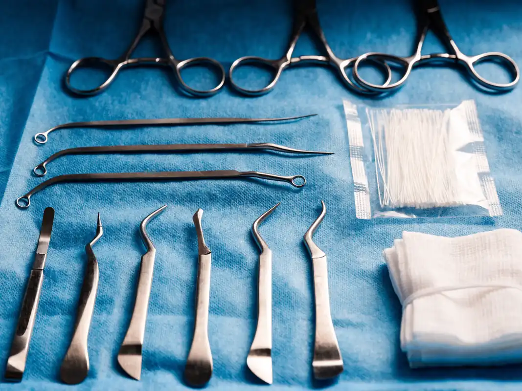
After diagnosis and consent, local anaesthetic is normally used. Pain control can be more difficult in a severely inflamed tooth, so tell the clinician if sensation persists. A protective rubber dam is commonly used to isolate the tooth and reduce contamination. The dentist opens the tooth, locates the canals, removes affected tissue, cleans and shapes the space, disinfects it and seals it with an endodontic filling material.
The number and length of appointments vary. Anatomy, active drainage, previous treatment, symptoms, restorability and the need for an interim dressing can all change the schedule. Treatment should not be sold as certain to finish in one appointment before examination.
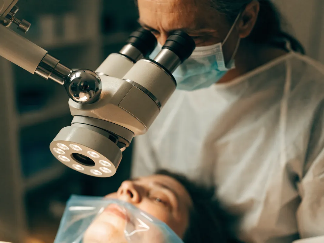
Magnification, electronic length measurement, mechanical instruments, irrigation methods and filling materials are technique choices. Ask what is planned and why, but do not treat a brand name as proof of quality or suitability. Important basics include diagnosis, isolation, working-length control, disinfection, a sound seal, adequate records and a realistic restoration plan.
Temporary material may be placed between stages or before the final restoration. Follow instructions about chewing and contact the provider if the temporary restoration is lost, the bite feels high or symptoms worsen.
Isolation is a safety and quality question
The operating field needs protection from saliva and oral bacteria, and the patient needs protection from instruments and irrigating solutions. Rubber-dam isolation is widely used for this purpose. Ask how the tooth will be isolated and what clinically justified alternative is planned if ordinary isolation is difficult.
Isolation is not a decorative photograph for marketing. It must work throughout the relevant procedure and around the actual tooth. Existing crowns, broken teeth or deep margins may require additional steps before a reliable seal can be established.
If a provider says isolation cannot be achieved, ask how contamination and patient protection will be managed and whether referral, restoration first, a different plan or extraction should be considered. The answer should be specific to the case.
Access and canal location preserve a difficult balance
The clinician must reach the pulp chamber and locate relevant canal anatomy while preserving enough tooth structure for later restoration. Too little access can obstruct cleaning; unnecessary removal can weaken the tooth. Existing crowns, posts, calcification and unusual anatomy can make this balance harder.
Ask whether old restorative work will remain, be accessed through or be removed. If treatment proceeds through a crown, understand the risk of damage, hidden leakage and later replacement. If a restoration is removed, clarify the temporary and definitive plan before travel.
Canals may be difficult to locate or negotiate. Magnification, illumination and appropriate instruments may help, but no device eliminates anatomical uncertainty. The record should identify canals treated and any anatomy that could not be managed as intended.
Working length and canal preparation need control
Cleaning requires an estimate and verification of how far instruments and irrigants should work within each canal. Electronic measurement and radiographs may contribute, but technique depends on anatomy and clinical judgement. A single screenshot or device name is not proof that the entire canal system was managed.
Preparation aims to create access for disinfection and filling while respecting root shape and strength. Curvature, narrow canals, calcification, previous errors and resorption increase difficulty. Ask how the operator will respond if a canal blocks, transports, perforates or cannot be negotiated safely.
The patient record should include relevant working information and deviations from plan. If treatment cannot proceed, consent should be renewed before referral, another attempt, surgery or extraction.
Irrigation and disinfection have real hazards
Mechanical instruments cannot touch every part of a complex canal system, so chemical cleaning is commonly part of treatment. The exact solution, concentration, volume, delivery and activation are clinical choices. They should follow current instructions and patient-specific safety considerations rather than a website recipe.
Ask how the tooth and soft tissues are protected, how delivery depth is controlled and how allergy or accidental exposure would be managed. Pain, swelling, bleeding or another unexpected event during irrigation requires direct clinical response and documentation.
No marketing phrase such as laser cleaning or deep sterilisation proves that all microorganisms have been removed. The useful comparison is the complete disinfection and sealing protocol, the operator's judgement and the contingency plan when drainage or symptoms persist.
One visit or several visits depends on findings
Appointment count can change with diagnosis, anatomy, drainage, symptoms, previous treatment, time available, patient tolerance and the need for an interim dressing. The NHS patient overview notes that treatment often involves more than one appointment, but that is not a rule for every tooth.
Ask what must be true before the canal is definitively filled and what would cause postponement. A timetable built around a flight should not override ongoing drainage, inability to dry the canal, unresolved anatomy or another clinical reason to wait.
If an interim dressing and temporary restoration are used, obtain written instructions, the expected review point and a plan if the seal is lost. A temporary stage is active treatment requiring follow-up, not permission to leave the tooth indefinitely.
Filling the canal is not the end of treatment
Once the clinician considers cleaning complete, the canal space is filled and the access is sealed. The material and technique should be recorded. Images can help document the result, but an image alone cannot show every branch, seal quality or future healing.
The tooth then needs an appropriate coronal restoration to reduce leakage and restore function. Responsibility may be split between an endodontic and restorative clinician, so the handover should state who provides the temporary seal, core, onlay, crown or other definitive restoration and on what clinical timeline.
Leaving a structurally weakened or temporarily sealed tooth without a definitive plan increases uncertainty. Before departing, know what can be chewed, what symptoms require contact and which clinician owns the next step.
The definitive restoration needs its own consent
Not every root-treated tooth needs the same restoration. Tooth position, remaining walls, crack risk, access shape, existing work, gum margins, bite and planned function influence whether a direct filling, onlay, crown or another restoration is considered. Root canal treatment does not automatically justify a crown, and a crown does not rescue an unrestorable root.
Ask what tooth tissue will be removed, which material category is proposed, how margins and cleanability will be managed, and whether a core or post is needed. A post can help retain a restoration in selected cases; it does not strengthen the root and adds its own removal and fracture considerations.
The permanent restoration, laboratory work, temporary protection, reviews and future maintenance should appear separately in the written plan and quote. Do not compare canal-treatment prices while ignoring the restoration that makes the tooth usable.
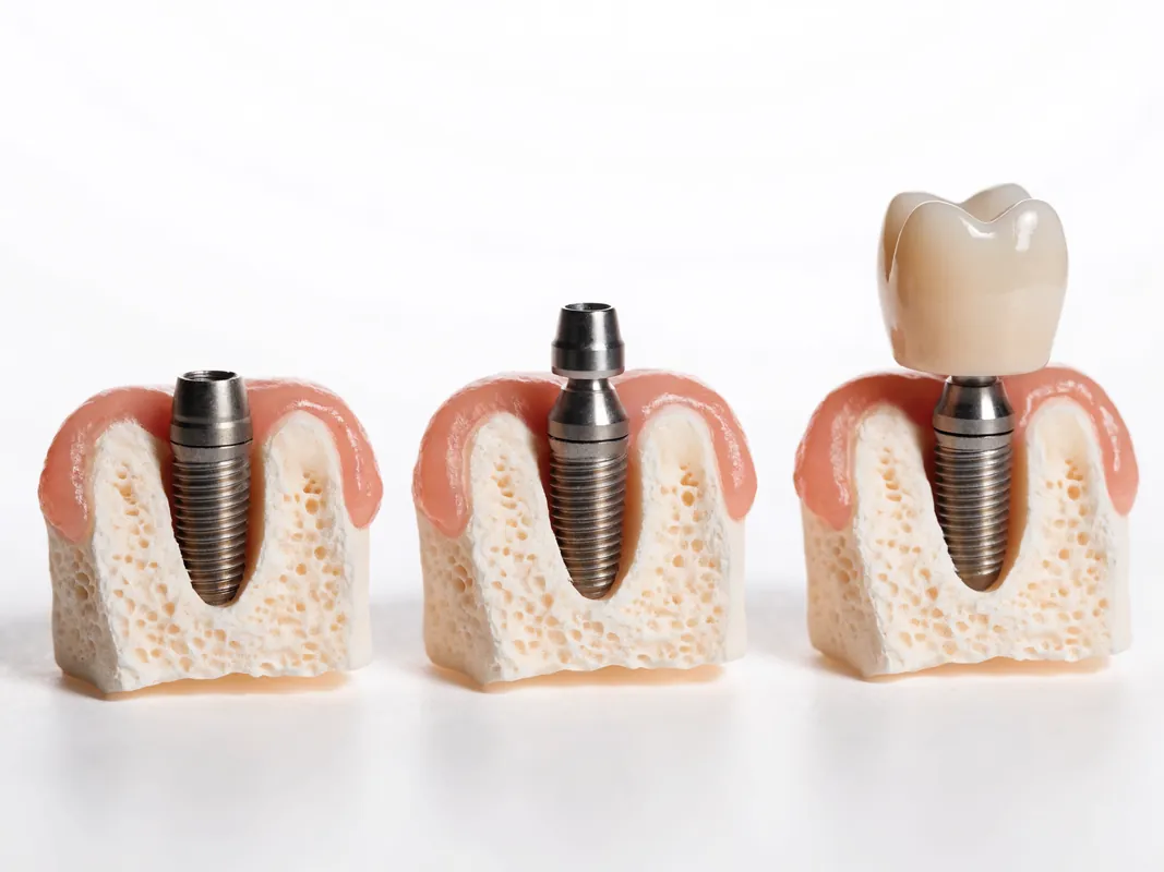
If a crown material is proposed, use the zirconia crowns service guide to structure material, preparation, laboratory and traceability questions without assuming zirconia is suitable. If extraction leads to an implant proposal, treat that as a new diagnosis and read the dental implants assessment guide.
Limitations, risks and uncertain outcomes
Root canal treatment aims to control disease and retain the tooth, but healing cannot be promised. Canals can be narrow, curved, calcified or difficult to locate. Instruments can separate, perforation can occur, infection may persist, a crack may extend, and the tooth or restoration may later fracture. Some treated teeth need re-treatment, surgery or extraction.
Pain, tenderness and jaw soreness can occur after treatment. Severe, increasing or persistent pain, visible swelling, an allergic reaction, a lost temporary restoration or the return of earlier symptoms needs professional review. Follow the clinician's medication instructions; do not assume that a medicine suitable for another person is safe for you.
The final restoration matters. A back tooth with substantial tissue loss often needs cuspal protection, while another tooth may be restored differently. The endodontic provider and restorative dentist should state who is responsible for the permanent restoration and when it should be placed. Avoid heavy chewing on an unrestored treated tooth as advised.
The [University Hospitals of Leicester root canal leaflet](https://www.uhleicester.nhs.uk/wp-content/uploads/2025/06/336_102022-Root-canal-treatment.pdf) lists practical uncertainties including discomfort, swelling, separated instruments, perforation, canals that cannot be fully located or negotiated, treatment failure and possible extraction. It is a patient-information source, not a prediction for an individual tooth.
A separated instrument is a finding, not a slogan
An instrument can separate inside a canal, particularly in difficult anatomy or re-treatment. Its importance depends on location, infection, stage of cleaning, canal anatomy, access and the risk of retrieval or bypass. Automatic removal is not always the safest option, and automatic dismissal is not adequate either.
Ask for the event to be explained and documented, with an image where appropriate, the options, risks and operator or referral plan. Possible paths can include monitoring, bypass, retrieval, continued treatment, surgery or extraction after case-specific assessment.
The patient should receive the updated record and understand whether the event changes prognosis, restoration timing, follow-up or cost. Consent for the original procedure does not remove the need for a new discussion after a material complication.
Perforation and procedural damage require disclosure
A perforation is an unintended communication between the canal space and surrounding tissues. It can arise from disease, resorption or procedural access and instrumentation. Location, size, contamination, timing and repair access affect management.
If suspected, pause for assessment. Ask who identified it, what evidence supports the finding, whether repair or referral is considered, which material is proposed under current instructions and what alternatives exist. The record should distinguish a pre-existing defect from a procedural event where possible.
Do not let travel pressure convert uncertain damage into immediate definitive work without adequate explanation. Local follow-up may be necessary even when repair is attempted abroad.
Missed anatomy and persistent disease need a cause analysis
Symptoms or a lesion after treatment do not automatically prove negligence or require extraction. Possible contributors include missed anatomy, leakage, new decay, an inadequate restoration, complex microbial persistence, fracture, periodontal disease or a non-dental source. The assessment should test plausible causes rather than simply repeat the original procedure.
Compare the quality and restorability of the whole tooth. Re-treatment may be reasonable when access and a durable final seal are achievable. Surgery may address selected disease near the root end. Extraction may be appropriate when the tooth is fractured or cannot be restored. Monitoring may be considered in selected uncertain findings under clinician review.
Ask what evidence would make the team change from one path to another and how healing will be assessed. A dramatic radiograph comparison should not substitute for clinical symptoms, restoration status and dated follow-up.
Vertical root fracture changes the pathway
A vertical root fracture can make retention impossible or highly uncertain depending on the tooth and fracture. It may be difficult to diagnose because symptoms and images can overlap with persistent endodontic or periodontal disease. A narrow deep gum pocket, recurrent swelling or a particular bone pattern may raise suspicion but does not alone settle the diagnosis.
Ask what direct and indirect evidence supports the conclusion and whether specialist assessment is appropriate. Repeating canal treatment does not repair a vertical fracture. If extraction is proposed, request the findings and replacement or no-replacement options separately.
Where diagnosis remains uncertain, the consent discussion should describe that uncertainty and the possible stopping point during exploratory work. Charging and temporary protection if the tooth cannot be saved should be agreed beforehand.
Healing is monitored, not assumed
Reduced pain is welcome but is not the only measure of healing. Clinical review can consider symptoms, tenderness, swelling, sinus tract, function and the restoration. Imaging may be used at a justified interval to assess changes around the root.
Ask when review is due, which clinician owns it and what findings would prompt further treatment. Cross-border care needs an agreed route for sending records and obtaining local examination without waiting for a reply from another country.
A treated tooth can develop new decay, restoration failure, fracture or recurrent disease later. Long-term maintenance therefore includes ordinary oral hygiene, risk-factor control, restoration review and access to the historical record.
Alternatives to discuss
The first question is whether the pulp and tooth can be preserved more conservatively. In selected cases with a vital pulp, caries control, a protective pulp procedure or vital pulp therapy may be considered after diagnosis. If the pulp is irreversibly damaged but the tooth is restorable, root canal treatment may be an option.
Extraction is the main alternative when the tooth cannot or should not be retained. The consequences include a space and possible changes to chewing; replacement choices may include no replacement, a removable option, a bridge or an implant after separate assessment. Each path has different biological, maintenance, time and cost implications.
Doing nothing is also a decision, but an infected or painful tooth may worsen and may eventually require urgent care. Ask for the advantages, limitations and likely next steps of each reasonable option in your case.
Re-treatment and surgical options
Symptoms or disease can recur after previous treatment. Assessment should look for a missed canal, leakage, new decay, a defective restoration, complex anatomy or fracture. Non-surgical re-treatment may be possible, but existing posts and restorations can add risk and cost. Endodontic surgery may be considered in selected cases. Extraction may be more appropriate when the tooth is not restorable. A remote image cannot settle this balance.
Ask whether old restorative work must be removed, who replaces it and what happens if a crack or other stopping condition is discovered during treatment. Those possibilities belong in consent and in the quote.
Vital pulp treatment may preserve living tissue
In selected teeth, a biologically based pulp procedure may be considered instead of complete root canal treatment. Suitability depends on diagnosis, symptoms, exposure, contamination control, restorability, patient factors and the clinician's ability to review the tooth. It is not simply a cheaper or faster root canal.
Ask whether the pulp is thought to be vital, which findings support that view, what procedure is proposed, how the coronal seal will be achieved and how vitality or healing will be monitored. A later root canal may still become necessary if symptoms or disease develop.
If this alternative is rejected, request the tooth-specific reason. Age alone, travel convenience or a package timetable is not an adequate explanation without clinical context.
Extraction has immediate and later consequences
Extraction may remove a source of pain or infection when a tooth is not restorable or retention has poor value. It also creates a wound and, in many locations, a missing-tooth decision. Healing, appearance, chewing, neighbouring teeth, bone and future replacement choices can matter.
Compare no replacement, a removable option, a tooth-supported bridge and an implant only after the extraction and wider mouth are assessed. Each has different tissue effects, maintenance, timing, cost and failure modes. An implant is not an automatic equivalent to a natural tooth.
If extraction is the contingency during attempted root canal treatment, consent and pricing should cover that possibility before treatment begins. Ask whether immediate replacement is actually suitable or whether healing and later reassessment are safer.
Re-treatment, surgery and extraction need one comparison
Non-surgical re-treatment approaches the canal through the tooth. Endodontic surgery approaches selected disease around the root end. Extraction removes the tooth. Their feasibility depends on anatomy, previous materials, restoration, fracture, gum support, adjacent structures, medical factors and operator expertise.
Request a side-by-side explanation of objective, access, likely loss of tooth structure, restoration consequences, recovery, follow-up, material uncertainties and what happens if the chosen path cannot be completed. No single pathway is universally best.
An independent opinion may be valuable before removing a post, cutting through a complex restoration, performing surgery or extracting a strategically important tooth. Allow time for that opinion in a non-urgent case.
Consent includes meaningful stopping points
Consent is a continuing conversation, not one signature at the start. The patient needs the diagnosis, purpose, alternatives, material risks, limitations, costs and opportunity to ask questions. New findings during access, dismantling or canal negotiation can require a renewed decision.
Agree in advance which events trigger a pause: an unrestorable crack, inability to isolate, unexpected anatomy, perforation, a separated instrument, inability to negotiate a canal or a major change in the restoration plan. Name who can authorise a different procedure if the patient is sedated or temporarily unavailable to decide.
The right to stop does not erase work already performed, so the financial terms should explain completed stages, temporary protection and record release. Pressure from a deposit, flight or clinic schedule must not replace clinical consent.
A second opinion needs usable records
A useful second-opinion package includes symptoms and timeline, medical history, examination findings, pulp and apical diagnosis, restorability assessment, dated images, current restoration, prior treatment record and the specific question being asked. A cropped screenshot without date or orientation limits review.
Ask the second clinician to state what can and cannot be concluded remotely. Conflicting opinions may arise from different evidence, thresholds or assumptions. Resolve the factual differences rather than choosing the most confident sales message.
If a second opinion changes the plan, update consent, quote, operator, travel and aftercare arrangements before treatment. Preserve both records for later handover.
Your written quote checklist
Request a written treatment plan and written quote before booking non-urgent travel. The plan should identify the tooth, working diagnosis, proposed treatment, likely appointment range, operator, imaging, anaesthesia, temporary and permanent restoration, review and alternatives. It should distinguish confirmed items from those that depend on the clinical examination.
The quote should itemise assessment, radiographs, endodontic treatment, temporary filling, final restoration, medication if applicable, reviews and foreseeable additions such as re-treatment or specialist referral. It should name exclusions, cancellation terms and who is responsible if the tooth proves unrestorable. Travel-related expenses should remain separate unless individually described and priced.
A low headline fee is not a complete comparison if the crown or other definitive restoration, follow-up or management of complications is omitted.
Compare quotations tooth by tooth
The quote should use a clear tooth identifier and distinguish diagnosis from treatment. Confirm whether the amount covers primary treatment or re-treatment, removal of a restoration or post, imaging, temporary and definitive seal, core build-up, onlay or crown, laboratory work, reviews and complication management.
Ask which items are fixed, estimated or excluded until examination. Record the currency, payment stages, cancellation terms, refund rules and what happens if treatment stops because the tooth is unrestorable. A package total that cannot be mapped to clinical stages is difficult to compare or challenge.
Travel, hotel and transport services should be under separately identifiable terms. Their convenience must not make an unnecessary procedure appear clinically required or hide the cost of local contingency care.
Cross-border handover starts before treatment
Identify a dentist near home who can examine urgent symptoms, complete a restoration if agreed and review healing. Do not assume that an unfamiliar clinician will accept another provider's diagnosis, proprietary records or unfinished work without a new assessment.
The overseas and home clinicians need compatible records: tooth and diagnosis, procedure dates, canals treated, complications, temporary status, materials where relevant, images, definitive restoration, medication and review plan. Name who answers clinical questions after departure and the expected response route.
If the permanent restoration will be completed near home, obtain a written restoration prescription and protection instructions. If it is completed abroad, the home dentist still needs sufficient information for future maintenance.
Appointment and flight timing need clinical flexibility
Do not build an immediate journey to the airport around anaesthesia, sedation or an unresolved complication. Numbness, discomfort, jaw fatigue, medication effects and the need for review can affect safe travel and eating. The responsible clinician should advise for the actual case.
Leave capacity for the plan to change after examination. A canal may require another stage, a restoration may need laboratory time, symptoms may need review or an uncertain tooth may require referral. A rigid return ticket cannot make those steps unnecessary.
For a single stable tooth, compare the total burden of travel with competent care near home. Root canal treatment is often a poor stand-alone reason for a dental trip because continuity and prompt access matter.
Records, handover and aftercare
Keep copies of the clinical notes, diagnostic tests, pre- and post-treatment radiographs, tooth and canal details, materials record where available, consent, itemised invoice, restoration plan and aftercare instructions. Ask for a concise handover that a dentist near home can use.
Follow the written advice while numb and while a temporary restoration is present. Maintain gentle oral hygiene and avoid heavy biting on the treated tooth until the provider confirms it is restored. Arrange the definitive restoration and review rather than treating canal filling as the end of care.
Contact the treating provider about expected post-treatment symptoms. Seek local urgent care for severe or worsening pain, swelling, fever, difficulty swallowing or breathing, an allergic reaction, trauma or a lost temporary restoration that leaves the tooth exposed. International follow-up should supplement, not delay, accessible local care.
For an elective travel decision, compare the complete written pathway with treatment near home: assessment, canal treatment, permanent restoration, reviews and contingency care. For an urgent tooth, prompt local diagnosis is usually safer and better value than arranging a dental trip.
The [American Association of Endodontists post-treatment guidance](https://www.aae.org/patients/your-office-visit/post-treatment-care/) identifies concerns such as severe or persistent pressure, visible swelling, an allergic reaction, an uneven bite, loss of a temporary restoration and return of earlier symptoms. Use the treating clinician's case-specific instructions; an online list cannot determine whether a symptom is expected.
Medication advice must be individual
Pain relief and other medication depend on medical history, allergies, pregnancy, other medicines and current symptoms. Follow the named prescriber's written instructions and the medicine label. Do not exceed a dose, combine products unknowingly or copy another patient's regimen.
Antibiotics should not be promoted as routine insurance after treatment. Ask for the indication and what signs require review. A rash, hives, breathing difficulty, facial swelling or other suspected serious reaction needs urgent medical advice according to local emergency guidance.
If pain is not controlled or is increasing, contact a clinician rather than repeatedly escalating self-medication. The cause may be the bite, inflammation, infection, a lost seal, fracture or another problem requiring examination.
Eating, cleaning and temporary-restoration care
While numb, follow the clinician's advice to avoid injury from biting or hot food. If a temporary restoration is present, avoid loading it beyond the written instructions and report loss, fracture or a high bite. Continue oral hygiene carefully unless a clinician gives a specific temporary restriction.
A thin surface change can be different from complete loss of a temporary filling, but a patient cannot always judge the difference. Contact the provider and arrange local protection if the tooth is exposed. Do not place household material inside the access.
The permanent restoration and ordinary preventive care are part of tooth survival. Brush with fluoride toothpaste, clean between teeth as appropriate, control decay risk and attend reviews based on individual need.
Warning signs need a local route
Seek urgent local assessment for facial or neck swelling, fever with dental symptoms, difficulty swallowing or breathing, rapidly worsening pain, uncontrolled bleeding, significant trauma or signs of a serious medicine reaction. Contact the treating dentist promptly for severe or persistent pressure, a bite that feels markedly uneven, loss of a temporary restoration, new swelling or return of earlier symptoms.
Do not wait for an international coordinator to wake up if urgent care is accessible locally. Send the overseas provider an update and records afterwards, but prioritise direct clinical safety.
Before leaving the clinic, save the legal facility name, treating clinician, clinical phone route, written emergency advice and a record package that another dentist can understand. A social-media account is not an emergency care plan.
Long-term maintenance belongs in the original plan
A root-treated tooth still needs decay prevention, gum care, restoration checks and assessment if symptoms return. The restoration can wear, leak, fracture or develop recurrent decay. The tooth can also be affected by bite changes or new trauma.
Ask how healing and restoration integrity will be reviewed, whether images are clinically justified and which findings would prompt monitoring, repair, re-treatment, surgery or extraction. Avoid a universal review calendar copied from marketing; the interval should reflect diagnosis and risk.
Keep the record even after the tooth feels normal. Future clinicians may need to know canal treatment, materials, posts, restoration design, previous complications and image dates before another procedure.
Use the dental treatment timeline guide to separate clinical gates from travel dates, and the returning home after dental tourism guide to prepare a local handover. Neither page replaces case-specific advice.
Records to take home
Request the history and diagnosis, examination and pulp-test findings, restorability assessment, dated radiographs or other images, treatment dates, tooth and canals managed, isolation and complication notes, temporary status, canal and restorative materials where recorded, medication, consent, itemised invoice, definitive restoration, aftercare and review plan.
If there was a referral or laboratory stage, include the referral letter, prescription and supplied traceability record where relevant. If a material event occurred, request the updated explanation and image rather than an oral reassurance alone.
Store records securely and share health information through an appropriate channel. Ask the legal data controller about access, retention and correction; a booking agent is not automatically the clinical record holder.
Red flags in a root canal offer
Pause when an offer relies on:
- a final diagnosis from one image without symptoms or clinical tests;
- no restorability or crack assessment;
- an urgent infection delayed to fit a travel date;
- routine antibiotics presented as the treatment;
- no named clinician or verifiable professional registration;
- specialist language without recognised specialist status;
- one fixed appointment promise before examination;
- no isolation or contamination-control explanation;
- a device or brand used as proof of outcome;
- no permanent-restoration owner or price;
- no stopping rule if the tooth is unrestorable;
- no explanation of re-treatment, surgery, extraction or no-treatment alternatives;
- complications hidden from the patient record;
- a package fee that mixes clinical and travel services;
- no accessible local urgent-care or handover plan.
Resolve important gaps before payment or irreversible treatment. If several remain, obtain an independent opinion or choose a provider that supplies a complete, reviewable pathway.
Root canal decision worksheet
| Decision | Evidence to request |
|---|---|
| Urgency | Symptoms, examination and local emergency plan |
| Tooth identity | Clear tooth notation and complaint history |
| Diagnosis | Pulp and root-area diagnosis with supporting tests |
| Restorability | Remaining structure, crack, gum and bite assessment |
| Complexity | Anatomy, previous work, access and referral factors |
| Imaging | Prescriber, date, question and report where available |
| Operator | Named clinician, role and current registration |
| Isolation | Case-specific contamination and patient-protection plan |
| Endodontic stage | Access, canals, cleaning, filling and deviations |
| Restoration | Temporary and definitive owner, design and timing |
| Alternatives | Pulp preservation, root canal, extraction and no treatment |
| Complications | Stopping rules, disclosure, referral and contingency |
| Quote | Itemised clinical stages, exclusions and plan-change terms |
| Handover | Images, procedure record, materials and local care route |
| Review | Symptoms, restoration, healing evidence and named owner |
Sources and evidence boundaries
This guide uses the [NHS root canal overview](https://www.nhs.uk/tests-and-treatments/root-canal-treatment/), [NHS England dental care pathways guidance](https://www.england.nhs.uk/long-read/nhs-dentistry-care-pathways-guidance/), [NHS England restorative dentistry complexity standard](https://www.england.nhs.uk/wp-content/uploads/2022/10/B1640-clinical-standard-restorative-dentistry.pdf), [GDC consent standard](https://standards.gdc-uk.org/pages/principle3/principle3), [GDC record standard](https://standards.gdc-uk.org/pages/principle4/principle4), [AAE patient overview](https://www.aae.org/patients/root-canal-treatment/what-is-a-root-canal/), [AAE post-treatment guidance](https://www.aae.org/patients/your-office-visit/post-treatment-care/) and [University Hospitals of Leicester patient leaflet](https://www.uhleicester.nhs.uk/wp-content/uploads/2025/06/336_102022-Root-canal-treatment.pdf).
These sources explain general patient questions, professional records or selected UK care pathways. They do not diagnose an individual tooth, certify a Turkish provider, establish a universal technique or predict an outcome. Product instructions, professional status, evidence and regulations change; verify the current named clinician, facility, materials and jurisdiction before treatment.
Final decision rule
Treat urgent symptoms locally. For a stable tooth, choose the proposal that proves diagnosis, restorability, clinical ownership, isolation, complete endodontic and restorative stages, consent, records and accessible follow-up. Compare the whole tooth-retention pathway with competent care near home before treating a flight or package as part of the clinical decision.