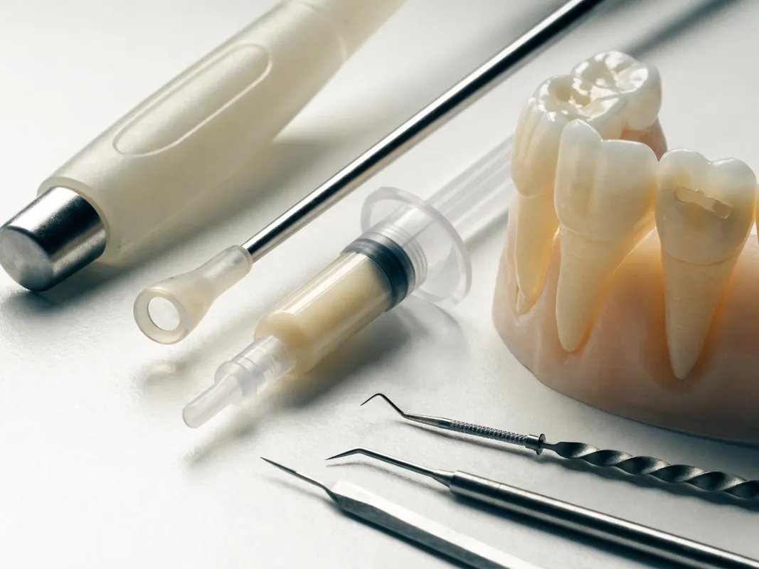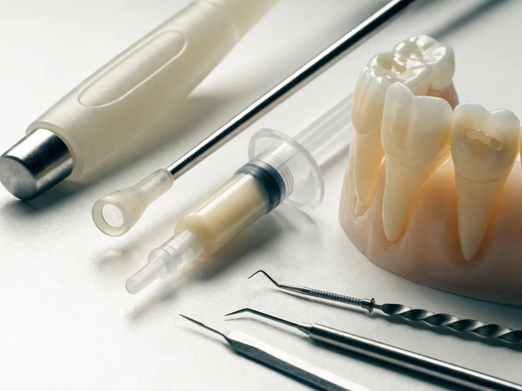
A diastema is a space between neighbouring teeth. The best-known example is a space between the upper central incisors, but spacing can occur anywhere in either arch and may involve one gap or many. A gap can be a healthy feature that a person values. It can also reflect normal dental development, tooth-size and arch-size difference, missing or small teeth, tooth position, periodontal migration, a supernumerary tooth, an oral habit, a frenum relationship or several factors together. Calling every gap a “veneers case” skips the diagnosis.
Closing a space changes more than the white area visible in a photograph. Orthodontics moves teeth and roots through bone. Direct composite adds material to tooth surfaces. A veneer covers part of a tooth and may require irreversible preparation. Frenum surgery changes soft tissue but does not by itself redistribute tooth size or move roots. Each option creates different effects on proportions, contacts, gums, speech, bite, cleanability, retention and future maintenance.
This is an evidence-led decision guide for adults researching gap-teeth treatment, including people considering dental care abroad. It is not a personal diagnosis, prescription, fixed treatment sequence, quotation, timetable, material selection or result promise. A named dentist must examine the mouth, identify the cause and health status, discuss no treatment and reasonable alternatives, explain uncertainty, obtain continuing consent and remain responsible for care. Orthodontic, periodontal, restorative, paediatric or other input may be needed. Photographs can organise questions but cannot establish the final plan.
A gap is a description, not a diagnosis
Begin by mapping the space rather than naming a treatment. Is it exactly between the upper central incisors, between a central and lateral incisor, distributed across several anterior teeth, beside a missing tooth, or part of generalised spacing? Is the upper dental midline aligned with the face and lower midline? Are the crowns upright, tilted or rotated? Are the roots parallel, divergent or convergent? Are adjacent teeth small, peg-shaped, restored, worn or displaced? Is the gap stable, closing, widening or newly visible?
The same width measured between crown edges can represent different biology. Two small central incisors in a broad arch are not the same as normal-width teeth with divergent roots. A space beside a congenitally missing lateral incisor is not the same as an isolated central diastema. A new gap associated with mobile teeth and gum disease is not an aesthetic design problem. A child in mixed dentition is not an adult restorative case.
A useful diagnostic statement includes the site, distribution, dentition stage, tooth and arch proportions, root position, periodontal condition, occlusion, likely causes, symptoms, patient preference and degree of certainty. “Two-millimetre gap” alone is not a diagnosis and cannot determine the number of restorations.
New or increasing spacing needs clinical assessment
A long-standing stable space without disease is usually not urgent. A newly appearing or widening space can be more significant, especially when accompanied by bleeding gums, recession, mobility, pain, swelling, a bad taste, trauma, a loose restoration or changes in the bite. Periodontal attachment loss and pathologic tooth migration can create or enlarge anterior spaces. Treating the visible gap with composite or veneers without stabilising disease can hide progression and complicate cleaning.
Seek timely dental assessment after trauma, for a tooth that changes position or colour, or where a space opens beside a tooth that feels mobile. Severe pain, spreading swelling, uncontrolled bleeding, difficulty breathing or swallowing, facial or neck swelling that is increasing quickly, collapse or another medical emergency requires urgent local care. Do not wait for a remote aesthetic consultation.
Cosmetic treatment cannot replace periodontal therapy, caries care, endodontic diagnosis, management of a fracture or assessment of an unerupted or supernumerary tooth. Antibiotics do not close a space and do not replace definitive dental care.
Age and dental development change the meaning of a midline gap
A maxillary midline diastema can be a normal feature of growth in primary and mixed dentition. As permanent incisors and later canines erupt, the position of the crowns and roots can change and some spaces reduce without restorative intervention. The American Academy of Pediatric Dentistry guidance on the developing dentition stresses diagnosis by dentition stage, eruption, oral habits, missing or supernumerary teeth and space relationships rather than a cosmetic shortcut.
An adult-focused marketing page should never be used to prescribe veneers or frenectomy for a child. Paediatric and orthodontic assessment may be needed when eruption is delayed, asymmetric or obstructed; when a permanent tooth is missing; when a supernumerary tooth or odontome is suspected; or when a habit or developing malocclusion is present. Radiographs are selected only when clinically justified, not as a routine photo-package requirement.
Development also matters for adolescents and young adults. Pulp size, eruption, gingival levels, tooth position and expectations can change. Removing sound enamel creates a restoration cycle that may continue for decades. Observation, orthodontics, additive composite or postponement may preserve more options. There is no age number on a website that automatically makes veneers appropriate.
Map the pattern of spacing across the whole dentition
An isolated upper midline gap draws attention, but planning must include the entire arch and bite. Record spaces between other teeth, tooth-size relationships, missing teeth, arch form, overjet, overbite, crossbite, rotations, incisor inclination and lower-arch alignment. A local restorative closure can make two teeth disproportionately wide while leaving the underlying distribution unchanged.
Useful records may include:
- a natural and broad smile, lip-at-rest and speech-related photographs;
- retracted frontal and side views;
- upper and lower occlusal views;
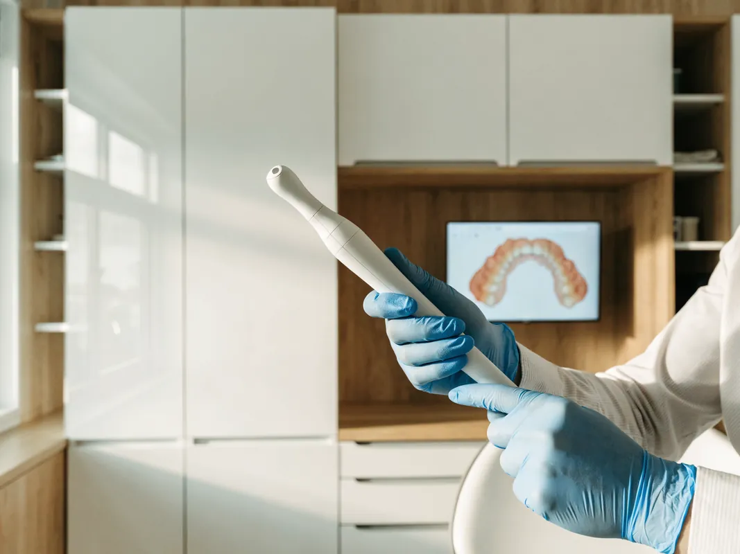
- scans or impressions where they add planning value;
- tooth-width and space measurements;
- periodontal charting and mobility where indicated;
- pulp tests for a symptomatic, traumatised or discoloured tooth where indicated;
- radiographs for a defined clinical question;
- a record of the bite and functional contacts;
- medical, medication, allergy and relevant habit history;
- the patient’s own description of what they want to preserve or change.
Measure tooth widths and available space rather than relying on a front-facing image. The goal is not to force every smile into a mathematical ratio. Measurements reveal whether closure by widening selected teeth would produce an acceptable transition or whether orthodontic redistribution, treatment of additional teeth or accepting some space deserves discussion.
Tooth size and arch size often explain generalised spacing
Spacing can result when the combined width of the teeth is relatively small for the available arch. Central or lateral incisors may be naturally narrow, peg-shaped or altered by wear. Several small differences across the arch can accumulate into visible spaces. Conversely, an apparently broad gap can result partly from tooth inclination or an arch relationship rather than small teeth.
The option should match the pattern. Additive composite may be able to reshape selected undersized teeth without preparation. Orthodontics may redistribute spaces so each restoration has a more maintainable width. A combination may move roots and crowns first, then add material where tooth-size discrepancy remains. Treating only the central incisors may be reasonable in one carefully assessed case and disproportionate in another.
Do not use a fixed “two, four, six or eight veneers” package as a substitute for a tooth-size analysis. The number of teeth should follow the natural smile, space distribution, restorative need, accepted transitions and tissue-preservation plan.
Missing, small, unerupted or supernumerary teeth require a different pathway
Congenitally missing lateral incisors, peg-shaped laterals, retained primary teeth, impacted teeth and supernumerary teeth can alter anterior spacing. A mesiodens or another obstruction may contribute to a persistent midline space or asymmetric eruption. These findings can require paediatric, orthodontic, surgical, restorative or prosthodontic assessment rather than immediate cosmetic coverage.
For a missing lateral incisor, the decision may include orthodontic space closure, opening or maintaining space for replacement, reshaping neighbouring teeth, retaining a suitable primary tooth, resin-bonded replacement or another option after growth and diagnosis. Each route changes root position, gum architecture, tooth proportions, retention and future replacement needs. Veneering the visible teeth without establishing whether a tooth is absent, impacted or expected to erupt is unsafe planning.
A panoramic radiograph may sometimes contribute to assessment, but it is not automatically sufficient and is not required solely because a patient sends a smile photograph. The responsible clinician chooses imaging according to history, examination, age and the specific question, with exposure justified and findings documented.
Frenum appearance alone does not prove that surgery is needed
The upper labial frenum is the fold of tissue between the upper lip and gum. Certain papillary or papilla-penetrating attachments have been associated with maxillary midline diastema, but association is not the same as proof that the frenum is the only cause or that cutting it will close the gap. Examination should consider blanching or tension, papilla and periodontal findings, eruption stage, root position, tooth size, habits and other possible causes.
A 2022 systematic review found limited and heterogeneous evidence, advised ruling out other dental and oral causes and reported that current literature generally recommends against performing frenectomy before orthodontic management of the diastema. A retrospective study found closure was more predictable when frenectomy was combined with orthodontic treatment than after frenectomy alone. These findings do not create a universal sequence; they show why “five-minute frenectomy prevents recurrence” is an unsupported promise.
Frenectomy has potential discomfort, bleeding, scarring, altered tissue anatomy and healing considerations. Its indication, method and timing belong to an appropriately qualified clinician after diagnosis. It should not be added as a free cosmetic extra, and it should not be sold from a photograph.
Habits and tongue posture need careful, non-deterministic assessment
Thumb or finger sucking, lip habits, tongue posture and other oral behaviours can be associated with spacing and incisor position, particularly during development. The presence of a habit does not prove that it caused an adult’s gap, and a single photograph cannot diagnose tongue thrust. Speech, swallowing pattern, airway concerns, dental and skeletal relationships and duration of the behaviour may need broader assessment.
If a functional habit is relevant, discuss whether it affects stability and whether referral or behaviour support is appropriate. Do not promise that a veneer will resist every force or that an exercise will permanently close a space. Restorations can chip, debond or wear; orthodontically moved teeth can relapse; untreated disease can progress. Stability planning must be specific to the diagnosed mechanism.
Periodontal health is a gate before aesthetic closure
Bleeding, inflammation, attachment loss, recession and tooth mobility change the decision. Periodontal disease can cause pathologic migration, flaring and new spaces. Closing contacts without treating disease can make plaque control harder and may create over-contoured restorations around unstable tissues. Gingival levels can also change after inflammation is controlled, altering proposed margins and tooth proportions.
The periodontal baseline should record plaque control, bleeding, probing findings, recession, mobility and any indicated imaging. Stabilisation and a maintainable supportive-care plan come before elective additive or indirect restoration. Aesthetic closure should not imply that gum disease is cured.
Where the papilla does not fully occupy the space, the patient should understand that closing the incisal gap may not eliminate a dark gingival embrasure. Contact position, root proximity, bone and tissue architecture influence papilla appearance. Trying to fill every black triangle with bulky material can compromise cleanability and contour.
Define the patient’s goal without treating natural variation as a defect
Some people value a midline diastema as part of their identity, culture or family appearance. Research on smile perception shows that preferences are not universal. A gap is not disease simply because a marketing image removes it. No treatment is a legitimate option when oral health is stable and the person does not want change.
Ask what the patient notices: the width of one space, generalised spacing, tooth shape, colour, asymmetry, a black triangle, speech, food trapping or something else. Ask whether partial closure is acceptable, whether the midline gap should remain smaller rather than disappear, and which natural features must be preserved. Show the smile at conversational distance as well as close-up.
The consent discussion should distinguish a health indication from an aesthetic preference. A person may choose monitoring, accept the space, defer until orthodontic information is available or stop after a diagnostic mock-up. Declining treatment is not a failed consultation.
Use a tooth-preservation option ladder
A conservative option ladder prevents a quick jump from “gap” to irreversible coverage:
- No treatment, reassurance, monitoring and routine prevention.
- Treat disease or a structural problem that is unrelated to cosmetic closure.
- Observe development or eruption where appropriate.
- Address an active habit or functional factor where clinically relevant.
- Orthodontic assessment to close or redistribute space and align roots.
- Additive direct composite to reshape selected suitable teeth.
- Combined orthodontic and additive restorative treatment.
- Indirect veneers only when the expected benefit justifies preparation, maintenance and future replacement.
- Crowns only where structural or restorative findings independently justify full coverage.
- Appropriately indicated periodontal or frenum treatment as part of a cause-led interdisciplinary plan.
Not every patient must try every step. The clinician should explain why a less destructive option is unsuitable or insufficient for this case. Cost, travel and an event date are relevant preferences, but they do not turn an irreversible option into a clinical necessity.
Orthodontics changes position rather than widening teeth
Orthodontic treatment can close a space, redistribute spaces, align dental midlines, change incisor inclination and improve root position. It may be especially relevant when teeth are rotated, flared or crowded elsewhere; when several spaces need redistribution; when a missing or small tooth affects proportions; or when widening central incisors would be visually or biologically undesirable.
Orthodontic assessment considers the whole bite, periodontal health, roots, eruption, skeletal relationships and retention. Clear aligners, fixed appliances and other methods are not interchangeable products that can be selected from a web page. Suitability, attachments, enamel reduction between teeth, extractions, anchorage, refinements and monitoring are clinician-owned decisions. A treatment-duration estimate must remain individual and can change.
Orthodontics does not change intrinsic tooth size or colour. After movement, small teeth may still benefit from additive composite or another restoration. This is why combined planning can be more conservative than making untouched but widely spaced teeth substantially broader with veneers.
Retention is part of orthodontic consent, not an afterthought
Teeth can move after orthodontic treatment, and spaces can reopen. NHS and orthodontic patient guidance describe retainers as an important part of maintaining the corrected position. The retainer type, wear plan, monitoring, replacement and repair route should be documented by the orthodontic provider. There is no universal wear schedule that a general web page can prescribe.
If composite is added after orthodontic space redistribution, the final restoration can alter the fit of a removable retainer. Coordination is needed so scans, bonding and retainer delivery follow a safe sequence. A fixed retainer may have hygiene, breakage and monitoring implications. The patient needs a local plan for a lost, distorted or detached retainer.
Frenectomy is not a substitute for retention. Nor does a bonded restoration guarantee that neighbouring teeth cannot move. The diagnosed cause, root position, occlusion, periodontal support and adherence to retention all affect stability.
Direct composite can be an additive, repairable option
Direct composite can add width to selected teeth, reshape peg-shaped or undersized teeth and close or reduce spaces without laboratory fabrication. In suitable cases, it may require little or no removal of sound tooth tissue. It can also be repaired, polished, modified or removed more readily than an indirect ceramic restoration, although removal without affecting enamel is not always perfectly reversible.
Clinical studies of direct composite for anterior space closure report useful performance in selected populations, but they do not promise a lifespan for an individual. A randomised trial comparing direct composite and indirect ceramic veneers for multiple diastema closure found no statistically significant survival difference during its two-year follow-up. Other observational studies report maintenance, colour, surface, chipping and repair findings over longer periods. Different operators, materials, cases and outcome definitions limit direct comparison.
Composite may stain, lose gloss, change contour, chip, fracture, debond, wear or need repair. Contact design and isolation can be demanding. The decision should consider enamel, gap distribution, tooth proportions, bite, habits, colour goal, maintenance access and the clinician’s documented plan—not a slogan that composite is temporary or ceramic is permanent.
Veneers are restorations, not orthodontics without braces
A veneer changes the visible form of a tooth by covering part of its surface. It can widen a tooth and reduce a visible space, but it does not move the root, treat periodontal migration, remove an obstruction, replace a missing tooth or correct the entire bite. Describing veneers as “closing a gap without moving teeth” is incomplete unless the resulting proportions, contacts, gums, function and tissue cost are also discussed.
Veneer treatment can require removal of sound enamel and is irreversible once preparation has occurred. Risks include sensitivity, dentine exposure, pulp effects, fracture, chipping, debonding, marginal stain, caries, gum recession, contour problems, colour mismatch, repair and future replacement. Preparation, bonding and long-term behaviour differ when margins extend into dentine or existing restorative material. Enamel preservation is an important planning objective.
An indirect veneer may be reasonable after diagnosis when less destructive options do not meet an informed goal, the proposed width and contour are maintainable, the substrate and bite are suitable, and the patient accepts the restoration cycle. It is not automatically superior because the gap has a particular millimetre width.
For a broader overview of irreversible restoration planning, see dental veneers and the whitening versus veneers decision guide.
Crowns need a structural indication, not a spacing target
A crown covers more tooth structure than a veneer and should not be proposed for healthy anterior teeth merely to close a space quickly or create opacity. Structural breakdown, extensive existing restorations, cracks, endodontic treatment or another tooth-specific finding may influence coverage, but the indication must be documented for each tooth.
Using crowns to make several sound teeth wider exposes them to greater preparation and future restorative cycles. Ask which exact structural finding makes a crown necessary, what alternatives were considered, how much tissue is expected to remain and how the pulp risk is managed. “Smile makeover” and “full coverage looks stronger” are not diagnoses.

Space closure must respect tooth proportions and transitions
Closing a midline space by adding equal width to two central incisors may appear simple on a flat image. In three dimensions, added width changes the apparent ratio of width to height, line angles, contact length, embrasures, facial curvature and transition to the lateral incisors. A result can close the dark line yet make central incisors look blocky or create abrupt steps beside adjacent teeth.
The design should compare several distributions: leaving some space, adding to two teeth, adding smaller amounts across more teeth, moving teeth first, or combining movement with additive restoration. The patient should see why each option changes the amount of natural tissue treated. Mathematical smile ratios can support discussion but should not be imposed as universal beauty rules.
If more teeth are proposed, the added benefit and tissue cost of each tooth must be explicit. Treating eight healthy teeth simply to avoid a visible transition may not be proportionate when orthodontics or selective composite could solve the underlying size distribution.
Contact areas, papillae and cleanability are biologic design constraints
The contact between anterior teeth is an area, not a single dot. Its position and length influence the visible embrasure, food passage and papilla appearance. The roots, bone crest and soft tissue help determine whether the papilla fills the gingival space. Restorations cannot guarantee complete papilla fill.
An overlong or over-bulky contact may hide darkness but create an unnatural square appearance or make interdental cleaning difficult. A short incisal contact may leave a black triangle. A deep subgingival contour can inflame tissue or complicate plaque control. The clinician and laboratory should document the intended contact and emergence profile and check whether floss or an appropriate interdental method can pass as advised.
Inflamed gums should be stabilised before final margins and papilla expectations are judged. If gum contouring is proposed, diagnose why the margin differs and read the veneers and gum-contouring guide before consenting to combined procedures.
A diagnostic wax-up and mock-up test shape, not biology or longevity
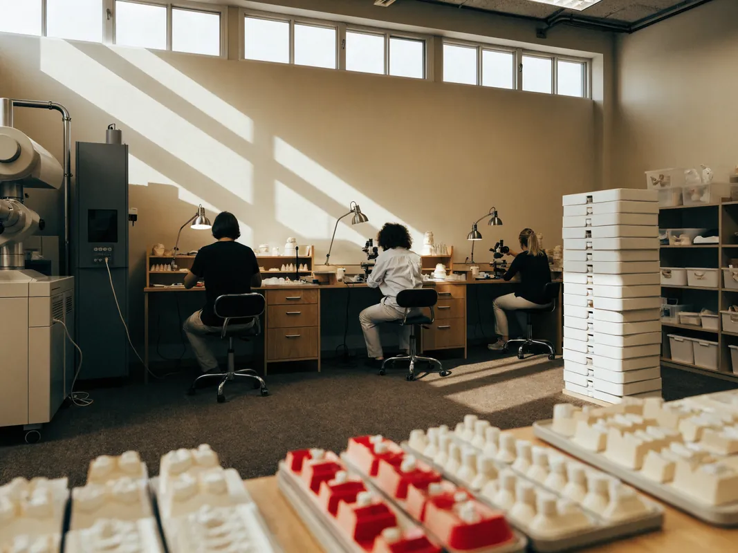
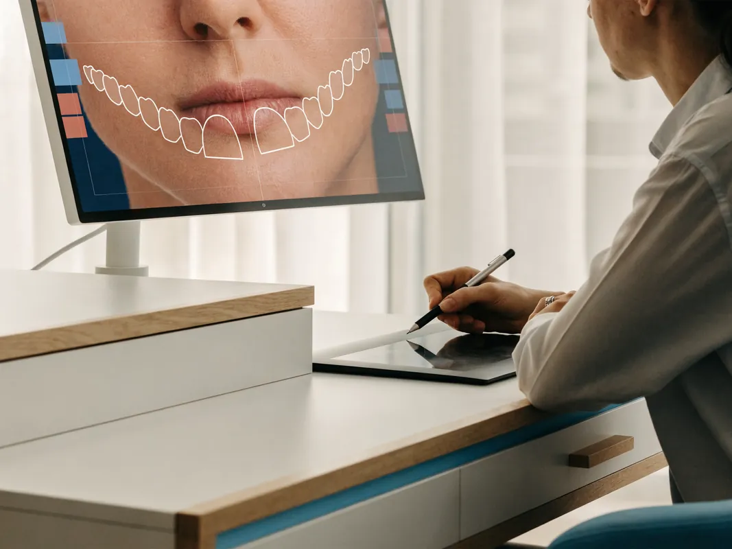
A wax-up or digital design can explore space distribution, tooth width, length, line angles and transitions. A physical mock-up placed over unprepared teeth can help the patient assess broad shape, lip support, speech and smile display. It is particularly useful when the alternative is to widen several teeth or change incisal length.
The mock-up has limits. Material placed over unprepared teeth can be bulkier than the intended restoration. It does not reproduce final optical properties, margin fit, bonded colour, gingival response or long-term function. A screen preview can be altered by lens, lighting, calibration and software. It is a decision aid, not a result guarantee.
Record what the patient accepts and rejects: full closure or residual space, width, length, symmetry, edge position, speech sounds and the view at conversational distance. If the mock-up only works by adding unacceptable bulk, the plan should change rather than automatically remove more enamel.
Additive design does not always mean no preparation
Some composite and veneer designs can be largely additive, but a website cannot promise “no-prep” or a fixed preparation depth. Tooth rotation, facial prominence, existing restorations, caries, colour, material thickness, margin placement and the selected contour may require modification. Conversely, a proposed preparation must not be justified merely because it is routine.
Ask for a preparation plan by tooth and surface. The preoperative scan, mock-up or reduction guide can help document intended tissue removal. After preparation, photographs or scans should record the actual substrate, enamel and dentine exposure and preparation colour. If the plan changes from additive composite to indirect veneers or from veneers to crowns, the clinician must explain why and obtain renewed consent before proceeding.
Preserving enamel supports bonding and retains future options. It does not remove every risk and cannot be reduced to an online percentage promise.
Material selection is case-specific and must remain traceable
Composite resins, glass ceramics, feldspathic ceramics and other systems have different optical, mechanical, bonding, repair and thickness characteristics. No named product or material is universally best for diastema closure. Evidence comparing direct composite with ceramic in selected cases does not prove equivalence for every patient or establish a winner for all follow-up periods.
The written plan should identify the material, manufacturer and product after the clinician selects it; the adhesive, cement and surface-treatment system where relevant; the laboratory and manufacture location for indirect work; and any applicable device documentation. The patient should receive enough information for maintenance and repair.
Terms such as “premium porcelain,” “Swiss quality,” “master ceramic” or “lifetime material” do not identify the system or prove suitability. Marketing adjectives are not a substitute for traceable records and case-specific reasoning.
Colour planning can change the number and sequence of restorations
If the patient also wants lighter teeth, whitening assessment may come before the final restorative shade decision. Whitening changes eligible natural teeth but not existing restorations in the same way. It can create sensitivity, mismatch or a need to reassess the endpoint. The treating dentist decides suitability, product, method, reviews and stopping criteria within the relevant legal framework.
Closing a gap with composite may be possible without changing colour. Treating additional healthy teeth solely to make everything brighter increases the restorative burden. If a patient is satisfied with natural colour, there is no need to add a whitening or veneer stage.
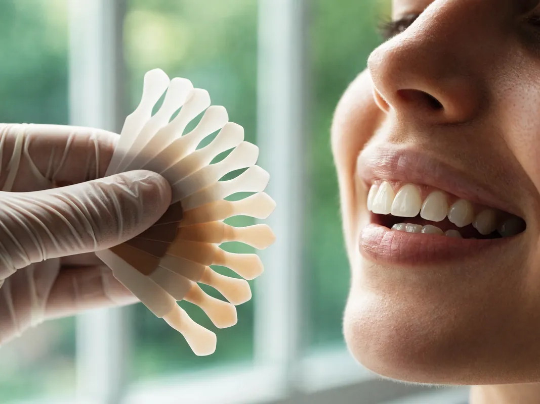
Photographs and shade devices support communication but are affected by lighting, dehydration, background and screen settings. A shade selected from a filtered selfie is not a clinical specification.
Bite, function and speech can veto an attractive front-view design
The added material occupies real space. The clinician should assess overbite, overjet, anterior guidance, edge-to-edge contacts, crossbite, protrusive and lateral movements, parafunction, wear and habits. A restoration that appears balanced from the front can contact heavily behind, interfere with movement or create a fragile edge.
Widening or lengthening anterior teeth can affect the tongue and some speech sounds. A mock-up or provisional may help identify a problem, but adaptation cannot be guaranteed or assigned a universal number of days. The patient should know who will review speech, bite and comfort if concerns persist.
Bruxism or other loading does not automatically exclude treatment, but it changes risk discussion, design, monitoring and any protective-appliance decision. A night guard is prescribed only after assessment and is not proof that a restoration cannot fracture.
Consent must remain active at every decision gate
Valid consent is an ongoing discussion, not a signature collected after travel or preparation. GDC Principle 3 describes explaining options and possible costs, answering questions and ensuring consent remains valid at each stage. These are useful patient-safety questions even where another jurisdiction governs the treatment.
Before irreversible work, the patient should understand:
- the diagnosis and uncertainty about cause;
- no treatment and monitoring;
- orthodontic, composite, combined and indirect options;
- what each option can and cannot change;
- whether sound enamel will be removed;
- the number of teeth and why each is included;
- contact, papilla, proportion, speech and bite limits;
- material, laboratory and maintenance implications;
- fracture, debonding, stain, caries, recession, sensitivity and replacement risks;
- retention or relapse considerations;
- costs, exclusions and change-of-plan terms;
- local aftercare and complaint routes;
- the right to pause, seek a second opinion or decline.
A deposit, booked flight or expiring offer must not override the right to stop. If the mock-up is unacceptable, gums are inflamed, a tooth is symptomatic, new information changes the cause or the proposed tissue removal increases, consent must be revisited.
Records should make the decision and future maintenance auditable
GDC Principle 4 describes complete records including history, findings, radiographs, photographs, models or scans, consent discussions, laboratory prescriptions and statements of conformity where applicable. A gap-closure record should include:
- the spacing map and tooth measurements;
- growth, eruption, missing-tooth and habit history where relevant;
- periodontal and occlusal findings;
- imaging justification and report where imaging is used;
- diagnostic alternatives and referrals;
- no-treatment, orthodontic, composite and veneer discussions;
- wax-up or digital-design versions;
- mock-up photographs and patient feedback;
- preparation and substrate records where relevant;
- material, adhesive, cement and laboratory identity;
- final contacts, contours, shade and occlusion;
- retainer or protective-appliance details if prescribed;
- consent, quotation, changes, reviews, repairs and aftercare.
Ask how records will be transferred to a local dentist. A social-media message and a before-and-after image are not a complete clinical record.
Demand an itemised written quotation
A package total hides whether the proposal is diagnostic, orthodontic, additive, surgical or indirect. Ask the legal provider for an itemised quotation separating:
- examination, photographs, scans and justified imaging;
- periodontal or caries treatment;
- orthodontic assessment, appliance, reviews, refinements and retainers;
- habit or other referral where relevant;
- composite trial, bonding, contouring, polishing and repairs by tooth;
- wax-up, mock-up and planning stages;
- each veneer or crown by tooth;
- preparation, provisionals, laboratory, try-in and bonding;
- frenum or periodontal treatment only if separately indicated;
- reviews, records and maintenance;
- complications, remakes and a changed plan;
- cancellation, postponement, refund and complaint terms;
- travel, accommodation and transfers as separate non-clinical items if purchased.
The quote should say what remains provisional until examination and what happens financially if treatment becomes less extensive, more extensive or unsuitable. “Free correction” may exclude travel, time off work, local care and biological problems. Read the written terms rather than assuming a warranty covers appearance, relapse or every repair.
A remote review can organise questions but cannot finalise closure
A remote enquiry can collect history, photographs, existing records and goals. It can flag that an orthodontic, periodontal or restorative assessment may be useful. It cannot palpate a frenum, measure periodontal attachment, test mobility, inspect every surface, assess roots, confirm eruption, diagnose an obstruction, reproduce colour or check dynamic occlusion.
Any remote plan, tooth count, timetable and quote must remain provisional. Before sending health information, identify the legal recipient, purpose, privacy notice, access and retention. Do not send unnecessary medical data through an insecure or unidentified account.
At the in-person examination, the responsible dentist must confirm diagnosis, health, alternatives, material, preparation, risks, records and costs. If a sales message promised two veneers and the examination supports orthodontics, monitoring or no treatment, the clinical plan should take priority.
Cross-border travel should follow clinical decision gates
The GDC guidance for patients considering dentistry abroad and the NHS treatment-abroad checklist advise checking the provider, treating clinician, qualifications, procedure, costs, insurance, records, follow-up and complaint arrangements. Gap closure may be elective, but it can still create irreversible preparation and long-term maintenance.
Before booking travel, obtain:
- the legal provider name and treatment address;
- the named dentist and current registration route;
- any named orthodontic or periodontal clinician and their role;
- the laboratory name and manufacture location for indirect work;
- a provisional sequence and the findings that could change it;
- enough contingency for assessment, trial, review or remanufacture;
- the local aftercare and urgent-care route;
- record-transfer, cancellation and complaint terms;
- insurance exclusions;
- separate written travel arrangements.
Do not allow a return flight to become a bonding deadline. A patient who rejects the shape, develops symptoms or needs further assessment must be able to pause. Travel convenience does not justify treating extra healthy teeth.
Local aftercare belongs in the initial plan
Identify a local dentist or orthodontic service before extensive treatment abroad. Ask whether they are willing to monitor the proposed work and what records they need. A local practice may not be able to reproduce an undocumented composite shade, ceramic system, contact shape or retainer immediately.
Maintenance can include periodontal review, caries prevention, cleaning around contacts, margin inspection, composite polishing or repair, veneer assessment, bite review and retainer monitoring. Composite and ceramic require material-compatible care. Natural teeth, gums and restorations can change differently over time.
Follow the individual provider’s cleaning advice. Do not force floss through a contact, use abrasive whitening products on restorations or adjust a retainer at home. Remote photographs can support communication but cannot replace necessary examination.
See caring for veneers long term for questions to take to the responsible dentist if indirect restorations remain under consideration.
Know the warning signs after treatment
Contact the responsible clinician for persistent or worsening pain, sensitivity that does not settle, a loose or fractured restoration, a sharp edge, a new bite interference, food trapping, bleeding or swollen gums, inability to clean a contact, a retainer that no longer seats, or a space that is reopening. Prompt local assessment may be safer than waiting for travel.
Seek urgent local care for severe pain with swelling, rapidly spreading facial or neck swelling, breathing or swallowing difficulty, uncontrolled bleeding, significant trauma or another emergency. These warnings do not predict a complication; they define when a web page is not enough.
Red flags before gap closure
Pause and seek an independent opinion if you encounter:
- a final diagnosis from a selfie;
- a fixed millimetre rule that automatically selects veneers;
- a claim that every frenum causes relapse or needs surgery;
- frenectomy sold before eruption and orthodontic assessment without explanation;
- periodontal mobility or new spacing ignored;
- an unerupted, missing or supernumerary tooth not investigated where relevant;
- orthodontics dismissed only because it takes longer;
- composite dismissed as short-lived without discussing repairability or evidence;
- veneers called permanent, reversible or preparation-free without case evidence;
- crowns proposed for healthy teeth solely to close space;
- a fixed two-, four-, eight- or twenty-tooth package;
- exact proportions or papilla fill guaranteed from software;
- no mock-up or change-control stage before preparation;
- no named dentist, legal provider or laboratory;
- an unitemised quote combining dentistry and travel;
- no retention, maintenance, record-transfer or local aftercare plan;
- pressure to bond because a flight or discount is approaching.
Questions to ask the named dentist and any orthodontic or periodontal clinician
- What is the diagnosis, not just the gap width?
- Is the space stable, developmental, pathologic or uncertain?
- Which teeth are small, missing, unerupted, displaced or restored?
- Are the roots and dental midlines appropriately positioned?
- Is periodontal disease or mobility contributing?
- Is imaging clinically justified, and what question will it answer?
- Is no treatment or monitoring reasonable?
- Could the space change with eruption or growth?
- Would orthodontics close or redistribute the space more conservatively?
- What retention would orthodontic treatment require?
- Could additive composite meet the goal?
- Can a composite trial show the proposed width and contact?
- Why is a veneer proposed for each named tooth?
- What structural finding would justify a crown?
- How much enamel is expected to remain?
- Is frenum treatment actually indicated, and when?
- Which other causes of the space were excluded?
- How will tooth widths and transitions be distributed?
- How will contacts, papillae and cleanability be managed?
- What residual space or black triangle cannot be ruled out?
- How will bite and speech be tested?
- What does the mock-up show, and what can it not show?
- Which material, adhesive or cement is proposed and why?
- Who is the legal provider and named treating clinician?
- Which laboratory will make any indirect restoration?
- What records and device documents will I receive?
- What exactly is included and excluded from the quote?
- What happens if the in-person diagnosis changes the plan?
- Who provides repairs, retention checks and urgent care locally?
- Can I pause or take the records for a second opinion before preparation?
A patient decision checklist
Before irreversible gap closure, confirm:
- My gap has been diagnosed rather than measured from one photograph.
- Growth, eruption, missing or extra teeth and periodontal causes were considered where relevant.
- I understand that a gap can be healthy and no treatment is an option.
- I have compared orthodontics, additive composite, combined care and veneers.
- I know why each proposed tooth needs treatment.
- I understand root movement and tooth widening are different interventions.
- I have seen a proportion and contact plan, not only a front-view render.
- I understand papilla fill and exact symmetry cannot be guaranteed.
- I know whether enamel will be removed and have preparation records planned.
- Frenum surgery is not being used as an automatic relapse promise.
- Retention, maintenance and local aftercare are documented.
- The material, laboratory and responsible clinicians are identifiable.
- The quote is itemised and travel is separate from clinical care.
- I can stop after the diagnostic or mock-up stage.
- I have enough contingency to avoid a flight-driven irreversible decision.
Evidence and further reading
These sources support the diagnostic and decision framework. They do not diagnose an individual, prescribe a treatment or guarantee a result.
- American Academy of Pediatric Dentistry, Management of the Developing Dentition and Occlusion in Pediatric Dentistry, latest revision 2024 and accessed 29 August 2026: dentition stages, eruption, habits, missing and supernumerary teeth, diagnostic records and timing. https://www.aapd.org/research/oral-health-policies--recommendations/management-of-the-developing-dentition-and-occlusion-in-pediatric-dentistry/
- NHS, Braces, reviewed 29 June 2026 and accessed 29 August 2026: orthodontic indications, oral health, monitoring and retention. https://www.nhs.uk/tests-and-treatments/braces/
- Review of maxillary midline diastema management with emphasis on cause, accessed 29 August 2026: growth, diagnosis, combined options and retention. https://pubmed.ncbi.nlm.nih.gov/18767455/
- Systematic review of superior labial frenum and maxillary midline diastema, accessed 29 August 2026: association, evidence limits, eruption and sequencing cautions. https://pubmed.ncbi.nlm.nih.gov/35248905/
- Retrospective study of diastema closure after frenectomy with or without orthodontics, accessed 29 August 2026: greater predictability with combined management than frenectomy alone in the study population. https://pubmed.ncbi.nlm.nih.gov/24392496/
- Systematic review of midline-diastema causes in children, accessed 29 August 2026: limited evidence concerning supernumerary teeth, frenum morphology and other factors. https://pubmed.ncbi.nlm.nih.gov/34135623/
- Randomised clinical trial comparing direct composite and indirect ceramic veneers for multiple diastema closure, accessed 29 August 2026: two-year outcomes in a small selected population, not a universal material ranking. https://pubmed.ncbi.nlm.nih.gov/39063806/
- Four-year clinical evaluation of direct composite diastema closure and recontouring, accessed 29 August 2026: performance, failure and repair findings with observational-design limits. https://pubmed.ncbi.nlm.nih.gov/33354867/
- Multicentre study of anterior direct composite build-ups, accessed 29 August 2026: long-term survival, repairability and quality outcomes in its study population. https://pubmed.ncbi.nlm.nih.gov/33491402/
- Clinical evaluation of direct composite build-ups after orthodontic treatment, accessed 29 August 2026: combined orthodontic-restorative sequencing in a selected cohort. https://pubmed.ncbi.nlm.nih.gov/25802222/
- Systematic review of veneer bonding to different tooth substrates, accessed 29 August 2026: enamel preservation and limitations of laboratory evidence. https://pubmed.ncbi.nlm.nih.gov/39207840/
- Systematic review of porcelain laminate veneer survival and complications, accessed 29 August 2026: heterogeneous evidence, fracture, debonding and maintenance. https://pubmed.ncbi.nlm.nih.gov/33807504/
- General Dental Council Principle 3, Obtain valid consent, accessed 29 August 2026: reasonable options, costs, continuing consent and documented discussion. https://standards.gdc-uk.org/pages/principle3/principle3
- General Dental Council Principle 4, Maintain and protect patients’ information, accessed 29 August 2026: clinical, photographic, laboratory and consent records. https://standards.gdc-uk.org/pages/principle4/principle4
- General Dental Council, Going abroad for dental treatment, accessed 29 August 2026: provider verification, costs, records, aftercare and complaints. https://www.gdc-uk.org/standards-guidance/information-for-patients-public/going-abroad-for-dental-treatment
- NHS, Treatment abroad checklist, accessed 29 August 2026: provider, travel, insurance, follow-up and contingency questions. https://www.nhs.uk/using-the-nhs/healthcare-abroad/going-abroad-for-treatment/treatment-abroad-checklist/
- NHS, How to find an NHS dentist in an emergency, accessed 29 August 2026: urgent dental access and emergency boundaries. https://www.nhs.uk/nhs-services/dentists/how-to-find-an-nhs-dentist-in-an-emergency/
The evidence supports cause-led diagnosis, respect for normal development, a no-treatment option, orthodontic and additive-restorative comparison, caution with frenectomy, tooth preservation, documented retention and long-term maintenance. It does not support a fixed gap-width rule, a universal veneer count, an automatic frenectomy, a preparation promise or a guaranteed closure result.