“Veneers and gum contouring” sounds like one cosmetic package. Clinically, it is at least two separate decisions: whether the visible concern comes from the teeth, the gingiva, the lips, the jaws, inflammation or a combination; and whether any irreversible change to tooth or periodontal tissue is justified. A photograph can start that conversation, but it cannot establish the diagnosis, the safe tissue boundary or the final margin position.
This page is a decision guide, not a diagnosis or a treatment promise. It does not assume that visible gum is a disease, that every uneven margin needs surgery, or that veneers are necessary after periodontal reshaping. It does not prescribe a laser, gingivectomy, crown-lengthening technique, veneer material, number of veneers or universal healing interval. Those choices belong to a named, appropriately licensed dentist after an examination, periodontal measurements, a restorative assessment and whatever records are justified for the individual case.
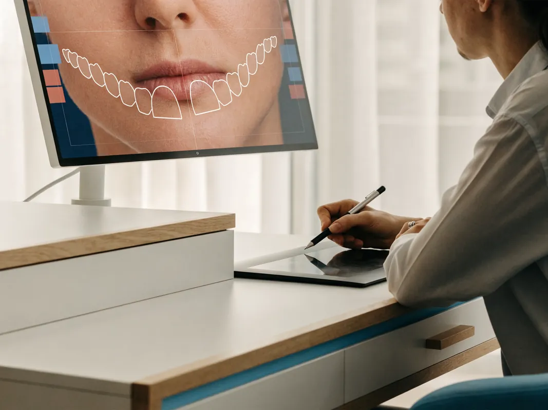
The central planning rule is simple: diagnose the reason for the display or asymmetry before changing either the gum frame or the teeth. The sequence should then protect health, tooth structure, function, cleanability and the patient’s right to reconsider. A digital preview can help people discuss proportions, but it is a communication record rather than a biological guarantee.
For the separate restorative fundamentals, see the dental veneers service guide. For design records and the difference between a preview and treatment, use the smile design guide. When inflammation or periodontitis is part of the presentation, the broader gum treatment and restoration sequencing guide explains why periodontal and restorative endpoints must be coordinated.
Start with the Complaint, Not the Procedure Name
Patients use phrases such as “gummy smile”, “short teeth”, “uneven gums”, “one tooth is lower”, “veneers look too square” or “my smile is not symmetrical”. These descriptions are useful because they identify what the person notices. They are not diagnoses. The same visible pattern can have different causes, and different causes can require completely different options.
The first consultation should record what is bothersome, when it is visible, whether it has changed, and what outcome the person would regard as acceptable. A person may dislike gum display only in a full animated smile but not at rest. Another may be concerned about one inflamed or receded margin. Someone else may mainly dislike tooth colour or wear and assume the gum is the problem. The planning team should not convert a subjective concern into a surgical indication without examining the anatomy and health.
It is also legitimate to choose no treatment. Gingival display is not automatically unhealthy, and symmetry measured on a static image is not the only definition of an attractive smile. The consent discussion should include observation, hygiene improvement, limited treatment and no-treatment options alongside any combined veneer-periodontal proposal.
“Gummy Smile” Is a Description with Several Possible Causes
The scientific term often used for a large amount of visible gingiva on smiling is excessive gingival display. The literature describes several possible contributors rather than one universal cause. A review indexed by PubMed discusses the importance of aetiology and diagnosis before choosing a treatment: [excessive gingival display—etiology, diagnosis and treatment modalities](https://pubmed.ncbi.nlm.nih.gov/19898712/).
Possible contributors include:
- Altered passive eruption. Part of the anatomical crown may remain covered by gingival tissue after eruption. The relationship between the gum margin, cemento-enamel junction and bone matters; a photograph alone cannot show all of those relationships.
- Dental or dentoalveolar position. Teeth may have erupted or moved in a way that changes incisor position, the gum line and the amount of display. Tooth wear can also trigger compensatory eruption and alter proportions.
- Lip length or mobility. A short upper lip at rest, a highly mobile lip during smiling, or both can influence the visible amount of gingiva. Removing gingiva does not correct every lip-related cause.
- Vertical jaw relationship. Skeletal proportions can contribute to display. A local gingival procedure cannot be assumed to correct a skeletal driver.
- Gingival enlargement or inflammation. Plaque-related inflammation, some medicines and certain systemic conditions can change tissue volume. The cause needs assessment rather than cosmetic trimming over active disease.
- Tooth shape, wear or restoration design. Naturally small crowns, worn incisal edges, existing restorations and uneven tooth dimensions can make the tissue frame appear dominant.
- Asymmetric recession or tissue loss. A margin may look “too high” because tissue has receded, not because the neighbouring gums need to be removed. Further subtraction may worsen the discrepancy.
- More than one factor. A person may have altered passive eruption, lip mobility, tooth wear and inflammation at the same time. A single-procedure label can conceal that complexity.
A systematic review comparing surgical and non-surgical approaches to excessive gingival display found substantial variation between interventions and called for more rigorous primary research: [Maleki and colleagues, 2024](https://pubmed.ncbi.nlm.nih.gov/38920855/). That evidence does not establish which option is right for an individual and should not be turned into a blanket sales claim.
Diagnose Asymmetry Rather Than Drawing a Symmetrical Line
Gingival asymmetry can arise from different tooth positions, different crown dimensions, recession, inflammation, altered eruption, previous restorations, trauma or normal facial asymmetry. A proposed line drawn across a smile photograph may be a helpful conversation aid, but it cannot decide how much tissue may be changed safely.
The assessment should distinguish the free gingival margin from the papillae between teeth. Lowering or raising a facial margin does not automatically rebuild a missing papilla or close a black triangle. Papilla form is influenced by the underlying contact, tooth shape, root position, periodontal support and interproximal anatomy. A plan that promises to “make every gum triangle identical” without examining those factors is not an adequate clinical plan.
It should also ask whether the apparent asymmetry is stable. Swollen tissue can change when plaque control improves. A provisional restoration can alter pressure and cleansability. Orthodontic movement can change both tooth and tissue positions. A margin should not be declared final while the condition that controls it remains unresolved.
Build a Shared Diagnostic Record Before Choosing Veneers or Surgery
A combined plan needs a diagnostic record that both the periodontal and restorative decisions can use. The exact record set is case-specific, but the following questions usually belong in the assessment:
- Medical and medicine history: Are there conditions, medicines, allergies, smoking or vaping, pregnancy considerations, bleeding risks, healing concerns or previous reactions that affect treatment choices?
- Dental history: Has the area been restored, traumatised, orthodontically moved, bleached, surgically treated or affected by recession before?
- Periodontal status: Are plaque, bleeding, probing depths, attachment levels, recession, tissue thickness and mobility documented where relevant?
- Caries, pulp and structural status: Are the proposed veneer teeth healthy, restorable and free from unresolved pain, cracks or active decay?
- Smile dynamics: What is visible at rest, during speech, in a posed smile and in a full animated smile? A single posed photograph can miss lip movement.
- Tooth and facial references: How do incisal edges, midlines, tooth axes, contact areas, lip line and facial asymmetry relate?
- Function: Is there wear, grinding, clenching, an unstable bite, restricted movement or a habit that could affect thin restorations or the periodontal tissues?
- Anatomical boundaries: Where are the cemento-enamel junction, bone crest, roots and proposed restorative margins? Direct measurement and justified imaging may be needed in selected cases.
- Patient priorities: Is the priority less display, better symmetry, lighter teeth, repaired wear, a less invasive option or simply understanding what is normal?
- Local aftercare: Who will review the tissues and restorations after the patient returns home, and who owns any complication pathway?
Photographs, scans, radiographs, periodontal charts, models and a mock-up can each answer different questions. They are not interchangeable. Imaging should be clinically justified and proportionate; it should not be ordered merely because a marketing workflow lists it.
Periodontal Health Is a Gate, Not a Cosmetic Detail
Elective contouring and veneer work should not be used to hide uncontrolled gingival inflammation or periodontitis. Redness, swelling, bleeding, suppuration, increasing mobility, recession or persistent bad taste may require diagnosis and disease management before an elective design is finalised. The NHS overview of [gum disease](https://www.nhs.uk/conditions/gum-disease/) explains that bleeding, swelling and bad breath can be signs that merit dental assessment.
Health does not mean that every patient needs periodontal surgery. It means the team should identify whether there is disease, establish an appropriate endpoint, and reassess whether the tissues are sufficiently stable and maintainable for the proposed restorative margins. Plaque control and daily interdental cleaning matter because veneers do not make the tooth-restoration interface immune to inflammation or decay.
The distinction between gingivitis, periodontitis, recession, altered passive eruption and purely aesthetic preference must remain visible in the record. They have different biological problems and different treatment choices. If a page or quotation simply says “gum contouring included” without a diagnosis, it is missing the reason for the procedure.
Respect the Supracrestal Tissue Attachment
Older explanations often use the term “biologic width”. Contemporary periodontal classification uses “supracrestal tissue attachment” for the epithelial and connective tissue attachment above the alveolar crest. The 2017 World Workshop introduced updated terminology and discusses periodontal phenotype and restorative-related conditions: [classification introduction and key changes](https://www.efp.org/fileadmin/uploads/efp/Documents/Campaigns/New_Classification/Reports/World_Workshop__Introduction__JCP_.pdf).
The practical meaning is not that one universal millimetre rule can be copied from the internet. It is that a restorative margin and a surgically repositioned gum margin must be planned in relation to the individual tooth, attachment, bone, tissue phenotype and cleansability. Removing only superficial tissue when the intended margin conflicts with the deeper anatomy can create instability or inflammation. Removing bone when it is not justified creates a different and irreversible intervention.
A periodontal assessment should therefore distinguish:
- tissue removal that changes the visible gingival margin without bone recontouring;
- a crown-lengthening procedure that may involve flap access and alteration of supporting bone;
- treatment of inflamed or enlarged tissue;
- management of recession or thin tissue where addition, protection or observation may be more appropriate than removal;
- restorative margin relocation or tooth-shape changes that could avoid periodontal subtraction;
- orthodontic or multidisciplinary options when tooth or skeletal position drives the display.
The technique should follow the diagnosis. “Laser gum contouring” describes an instrument or method, not the biological indication. A laser does not make every case flapless, eliminate the need to assess bone, or establish a universal healing interval.

Gingivectomy and Crown Lengthening Are Not Synonyms
A gingivectomy or gingivoplasty generally refers to removal or reshaping of gingival tissue. A clinical crown-lengthening operation may require a broader periodontal procedure, sometimes including bone recontouring, to expose more tooth structure while respecting the attachment apparatus. The American Academy of Periodontology’s patient information explains that crown lengthening can recontour gum tissue and sometimes bone: [What is crown lengthening?](https://home.perio.org/PRODUCTFILES/8118845/CrownLengthening_sample.pdf).
Which description applies cannot be decided from the desired appearance alone. The location of bone, the cemento-enamel junction, probing findings, phenotype, tooth proportions and restorative plan matter. The consent form and itemised quotation should name the proposed intervention accurately enough for the patient to understand whether tissue only, tissue and bone, or no periodontal surgery is planned.
Evidence on margin stability should be communicated cautiously. A systematic review of pre-restorative crown lengthening identified heterogeneous studies, high risk of bias and possible gingival margin rebound: [Pilalas, Tsalikis and Tatakis](https://pubmed.ncbi.nlm.nih.gov/27535216/). A later systematic review and meta-analysis also noted the limited evidence base when examining periodontal tissue stability: [Smith and colleagues](https://pubmed.ncbi.nlm.nih.gov/37251724/). These publications support reassessment; they do not supply a fixed timetable for every patient.
Periodontal Phenotype Changes the Risk Conversation
Periodontal phenotype includes tissue thickness and the broader supporting anatomy. A thin or recession-prone presentation raises different concerns from a thick tissue presentation. The clinician should consider whether subtraction may expose root surface, increase recession risk, alter papillae or create a margin that is hard to maintain.
Phenotype is not a label that guarantees a particular response. It is one part of risk assessment. Existing recession, prominent roots, a narrow band of keratinised tissue, previous surgery, inflammation and restorative margin position may all change the discussion. In some cases, soft-tissue augmentation, orthodontic movement, an additive restorative change or no treatment may be preferable to removing more tissue. The patient should be told when a desired outline conflicts with a safer biological boundary.
Compare the Additive, Subtractive and No-Treatment Options
The least destructive route that meets the person’s goals deserves explicit consideration. “Combined treatment” should not become an excuse to perform two irreversible procedures when one limited or reversible step could answer the concern.
No treatment or monitoring
If the tissues are healthy and the concern is aesthetic, observation is a valid option. Updated photographs or periodontal measurements can document whether an asymmetry is stable. The patient may decide that normal variation is acceptable after understanding the anatomy.
Hygiene and periodontal disease management
When inflammation or enlargement drives the appearance, cause-focused care and reassessment may change the tissue outline before any cosmetic decision. This is not cosmetic contouring; it is disease management.
Whitening or colour-only treatment
If colour is the main concern, supervised whitening may avoid veneers. Existing restorations do not whiten like natural tooth tissue, so sequencing and shade expectations still need discussion.
Additive composite or a diagnostic mock-up
Composite additions can test length, edge position or tooth width without immediately committing to laboratory veneers. Composite has its own maintenance and staining considerations, but it can help determine whether tooth addition alone changes the perceived gum-to-tooth proportion.
Orthodontic options
When tooth position, intrusion, extrusion, spacing or root alignment contributes, orthodontic assessment may change the gum line and contact relationships while preserving tooth structure. It may take more time and has its own burdens, but it should not be hidden merely because a faster restorative route is being marketed.
Limited enamel recontouring or edge repair
Small shape discrepancies may sometimes be addressed conservatively, subject to enamel thickness, function and sensitivity risk. Subtractive enamel recontouring is still irreversible and requires consent.
Veneers
Veneers can change visible tooth shape, colour, width and length. They do not correct every periodontal, lip or skeletal cause. The American Dental Association’s [veneer information](https://www.mouthhealthy.org/all-topics-a-z/veneers) notes that enamel may be removed and that the procedure may therefore be irreversible. Veneers also require ongoing care; decay and gum disease can still occur around restored teeth.
Periodontal reshaping or crown lengthening
This may be considered when the diagnosis and anatomy support a change in the gingival margin. The intervention must be named and its boundaries documented rather than sold as generic contouring.
Lip, muscle or jaw-directed options
Lip repositioning, muscle-directed injections and orthognathic approaches address different contributors and involve different clinicians, limitations and risks. They are not interchangeable with gingival removal. A 2024 systematic review found important differences and evidence limitations across surgical and non-surgical excessive-display interventions; it should not be used to claim one universally superior method.
Use a Tooth-Preservation Ladder Before Approving Veneers
A practical consent sequence starts with the least irreversible option and moves only as far as justified:
- clarify the concern and the no-treatment option;
- stabilise health and remove diagnostic uncertainty;
- test colour-only or additive alternatives where appropriate;
- use a mock-up or provisional stage to evaluate proportion, speech and function;
- define the minimum number of teeth and minimum preparation needed;
- reconsider whether veneers remain necessary after the tissue or tooth-position issue is addressed;
- document why any enamel removal and periodontal surgery are proportionate.
“No-prep” should not be treated as a guarantee of no tooth alteration. Contact areas, margins, emergence profile and space may still require change, and an overcontoured restoration can harm appearance, cleanability or tissue health. Conversely, aggressive preparation should not be normalised when an additive or orthodontic alternative could meet the goal. The named treating dentist should record the intended preparation approach and what would make it change.
Plan One Endpoint but Keep Two Clinical Decisions Visible
The gum frame and tooth design influence each other, so they should share an endpoint. That does not mean they should be sold or performed as one inseparable package. The record should show:
- the periodontal diagnosis and owner;
- the restorative diagnosis and owner;
- the proposed final gingival margins;
- the proposed incisal edges, tooth widths and contact areas;
- the anatomical limits that may prevent the previewed outline;
- the decision gates at which the plan can be changed or stopped;
- which stage is provisional and which is definitive;
- who reviews healing and who accepts the final restoration.
A diagnostic wax-up, digital design or intraoral mock-up can help relate tooth additions to the proposed tissue frame. A published clinical report describes a digital workflow for altered passive eruption and explains how combined data can support diagnosis and communication: [Pedrinaci and colleagues](https://pubmed.ncbi.nlm.nih.gov/37078683/). It is not proof that a digital guide makes every outcome exact, nor that every patient needs three-dimensional imaging.
When Gum Planning Usually Comes Before Definitive Veneer Margins
If the planned periodontal intervention is expected to change the visible facial margin, definitive veneer margin placement should ordinarily account for that change. Fabricating final veneers against a tissue outline that is intentionally going to move can create mismatched lengths, exposed edges, overcontour or compromised cleanability.
That principle does not create a universal appointment schedule. There may be a diagnostic mock-up before periodontal treatment, conservative preparation or provisional restorations during the process, and reassessment before final records. The appropriate sequence depends on disease status, the type and extent of periodontal procedure, tissue response, phenotype, provisional needs and functional findings.
The decision gate is clinical stability, not a date copied from a package page. The record should define what the clinician is waiting to observe: healthy tissue, acceptable plaque control, a stable margin on repeated review, comfortable cleaning, resolved inflammation, a confirmed restorative emergence profile or another case-specific endpoint.
When Restorative Planning Comes Before Any Gum Removal
Restorative planning should inform periodontal treatment even if the periodontal procedure is performed first. The proposed incisal edge, width-to-length relationship, contact position, papilla support, emergence profile and margin location determine whether removing tissue would actually improve the whole composition.
A mock-up may reveal that adding length to worn teeth corrects the perceived shortness without substantial gum removal. It may show that the proposed teeth would become disproportionately long if both crown lengthening and veneer lengthening were performed. It may also expose a functional or speech problem before irreversible work.
Restorative planning first does not mean final veneers first. It means defining a testable endpoint before tissue is removed. The patient should see which parts of a preview are cosmetic preferences and which are constrained by roots, bone, periodontal attachment and function.
Provisional and Final Restorations Have Different Jobs
A provisional restoration can protect prepared teeth, test contours, support communication and provide information about speech, bite, cleaning and appearance. It is not simply a cheaper version of the final veneer. Materials and durability differ, and provisional tissue contours may be deliberately adjusted during reassessment.
A final restoration is accepted after the relevant biological and restorative decision gates have been met. The patient should know whether a quoted “veneer” at a particular stage is a mock-up, direct temporary material, provisional laboratory restoration or definitive bonded restoration. Those words should not be used interchangeably.
The plan should also explain what happens if the tissue does not settle at the desired position, if the mock-up looks wrong, if the bite is uncomfortable, or if additional findings change the indication. Consent remains active throughout; an initial signature does not authorise every later change.
Healing Is a Clinical Process, Not a Universal Countdown
Periodontal tissues change through early wound healing, maturation and adaptation. The type and extent of intervention, tissue phenotype, plaque control, smoking, health, medicines, restorative contours and individual response can influence what the clinician observes. Research reports group averages, not a guaranteed personal timetable.
For this reason, the guide does not state that final veneers are always fitted after a fixed number of days, weeks or months. A responsible plan names the review criteria and allows the definitive stage to move if inflammation, margin change, sensitivity, provisional problems or cleaning difficulty remain. It should also avoid implying that a laser creates instant biological stability.
Patients travelling for care should be especially wary of an itinerary that leaves no meaningful reassessment gate between tissue alteration and an irreversible final restoration. A short trip can be convenient, but convenience is not a clinical endpoint. The no-travel option and a staged route with local care should be discussed.
Veneer Margin Design Must Protect Cleanability
The restorative margin should be placed only as deep as clinically necessary and must be compatible with the periodontal tissues. A hidden margin is not automatically a healthier or more aesthetic margin. Deep or bulky contours can be difficult to clean and may contribute to inflammation. Exposed or mismatched margins can create aesthetic concerns. The trade-off depends on substrate, colour, preparation, material, tissue position and the clinician’s ability to isolate and bond.
The proposed emergence profile—the way the restoration transitions from the tooth through the tissue zone—should be deliberate. An overbulked cervical veneer can compress or displace tissue and trap plaque. An undercontoured form may fail to support the intended appearance. Contacts and embrasures should permit cleaning rather than merely closing every visible space.
The patient should be shown how to clean around the provisional and final work. If the intended design cannot be cleaned with realistic daily measures, it is not a complete design.
Shade, Translucency and Surface Texture Belong in the Plan
Gum contouring does not solve shade mismatch, and very bright veneers do not solve an incorrect periodontal diagnosis. Shade planning should consider natural tooth colour, existing restorations, stump or substrate colour, material thickness, translucency, fluorescence, texture and lighting. A photograph on one screen is not a definitive shade record.
If whitening is being considered, the sequence should recognise that natural teeth may change shade while existing restorations do not. The written plan should identify which teeth are being treated, what happens to visible restorations outside the veneer set, and whether a mismatch is acceptable.
Laboratory communication should include the authorised tooth map, material, shade information, design records and approved changes. The patient should be able to distinguish a visual preference from a promise that ceramic will exactly copy a filtered simulation.
Function, Bite and Speech Can Override a Cosmetic Preview
Lengthening incisal edges or changing tooth guidance can affect speech and jaw movements. Thin veneers are still restorations exposed to functional load. Bruxism, clenching, edge-to-edge relationships, deep bite, crossbite, missing posterior support and parafunctional habits may change the choice of material, design, protective strategy or whether veneers are sensible at all.
A mock-up or provisional can be used to assess “f” and “v” sounds, “s” sounds, lip closure, comfort and functional contacts. Problems should be resolved before definitive bonding where possible. A beautiful static image does not compensate for a bite that feels wrong or a design that repeatedly chips.
The consent discussion should include the possibility of maintenance, repair or replacement. No restoration has an unlimited service life, and a combined periodontal-restorative procedure does not eliminate biological ageing, wear or future disease.
Risks Should Be Specific to Both Parts of the Plan
Potential periodontal concerns can include bleeding, swelling, discomfort, infection, delayed healing, sensitivity, recession, margin change, papilla alteration, root exposure, tissue colour change, asymmetry, need for further care and effects on adjacent sites. The relevant risks depend on the actual procedure; a consent form should not present every intervention as simple superficial trimming.
Potential veneer concerns can include sensitivity, enamel removal, bonding failure, fracture, chipping, staining at margins, colour mismatch, contour problems, gum irritation, decay, bite changes, need for repair or replacement and the possibility that a prepared tooth later needs additional care. The ADA advises patients to use a licensed dentist and notes that cavities can still develop under or around veneers.
Combined treatment adds coordination risks. A periodontal margin may change after the veneer has been designed. A provisional may irritate healing tissue. An altered contact may affect the papilla. A late plan change may alter cost, visits or the number of teeth. These interactions are a reason for explicit decision gates, not a reason to offer certainty about the combined result.
Consent and Change Control Must Stay Active
The UK General Dental Council’s [consent standard](https://standards.gdc-uk.org/pages/principle3/principle3) requires relevant options, risks, potential benefits and costs to be explained and treats consent as an ongoing process. It also states that changes to agreed treatment or estimated cost require renewed consent and documentation. These UK standards are useful questions for a UK patient to ask; they are not a statement that a provider abroad is regulated by the GDC.
Before irreversible treatment, the patient should receive plain-language answers to at least these points:
- What is the diagnosis for the gingival display or asymmetry?
- Which findings support tissue-only contouring, crown lengthening, a non-surgical alternative or no treatment?
- Which teeth are proposed for veneers, and why is each tooth included?
- How much tooth alteration is anticipated, and what could make it greater?
- Which stage is a mock-up, provisional or definitive restoration?
- What clinical findings must be present before final records or bonding?
- What are the material, margin and shade choices?
- Who is the legal treatment provider and named treating clinician for each stage?
- Who is responsible for aftercare and complications after travel?
- What changes require a new written quotation and renewed consent?
Consent is not satisfied by a before-and-after image, a package label or a signature obtained immediately before treatment. Patients need enough information and time to compare options, including the option to stop.
Records and Laboratory Traceability Make the Sequence Auditable
The GDC’s [record-keeping standard](https://standards.gdc-uk.org/pages/principle4/principle4) lists photographs, models, radiographs, consent documents and laboratory prescriptions among records where available. A coordinated case should preserve which record informed which decision.
Useful records may include:
- baseline periodontal charting and tissue photographs;
- smile photographs at rest and in function;
- tooth and restoration charting;
- justified radiographs or other imaging and their reports;
- scans, impressions, models, wax-ups and mock-up records;
- the approved gingival and incisal reference design;
- the actual periodontal procedure performed;
- preparation and provisional records;
- material and shade prescription;
- laboratory identity, prescription and relevant conformity documentation;
- bite records and adjustments;
- consent discussions, plan changes and patient approvals;
- postoperative instructions and local handover information.
The patient should ask how to obtain copies. Records are particularly important when periodontal treatment, restorative treatment, laboratory fabrication and aftercare involve different organisations or countries.

Demand an Itemised Written Quotation
A headline price for “veneers plus gum contouring” is not enough to compare plans. An itemised quotation should identify:
- the legal provider and location for each clinical stage;
- examinations and records included or charged separately;
- whether periodontal treatment means tissue reshaping, crown lengthening involving bone, disease treatment or another procedure;
- the teeth and surfaces involved;
- whether mock-ups, provisionals and definitive veneers are separate items;
- veneer material and laboratory information;
- medicines, imaging, sedation or anaesthesia where relevant;
- review visits and what is not included;
- what happens financially if the clinical plan changes;
- cancellation, postponement, remake, repair and refund terms;
- aftercare ownership after the patient returns home;
- travel, accommodation and transfer items only where explicitly stated in the written agreement.
Do not infer that flights, hotels, transfers, remedial travel, future maintenance or replacement are included. Compare the total clinical pathway and the exclusions, not just the number next to each veneer.
Plan Travel Around Decision Gates, Not Marketing Convenience
Dental travel adds distance between the patient and the treating provider. A combined periodontal-restorative plan may need reassessment, provisional modification, hygiene review or management of an unexpected finding. The travel plan should identify which stages genuinely need to be performed by the same team and which can safely be reviewed locally.
Before booking, ask for:
- the named legal provider and exact treatment location;
- the clinician responsible for periodontal diagnosis and procedure;
- the clinician responsible for veneer preparation and bonding;
- the planned order of records, tissue care, provisional and final stages;
- clinical criteria for postponing definitive work;
- a contact route during the trip and after returning home;
- the records supplied to a local dentist;
- written responsibility for complications, adjustments and remakes;
- the no-travel option and how it differs clinically or financially.
The GDC’s patient information on [going abroad for dental treatment](https://www.gdc-uk.org/standards-guidance/information-for-patients-public/going-abroad-for-dental-treatment) recommends researching qualifications, regulation, complaints, insurance, aftercare and the full cost. The NHS also provides a planning checklist for [medical treatment abroad](https://www.nhs.uk/using-the-nhs/healthcare-abroad/going-abroad-for-medical-treatment/). Neither resource can verify a particular provider; the patient must check the current facts.
Aftercare Protects Tissues and Restorations Together
Aftercare instructions should be individual to the periodontal intervention and the veneers. General themes include gentle plaque control as instructed, cleaning between teeth, following medicine directions, avoiding habits or loads the clinician has restricted, and attending planned reviews. The technique and timing of cleaning near a healing site should come from the treating clinician rather than a generic internet schedule.
Long-term care should examine both tissue health and restoration condition. Reviews may assess plaque, bleeding, recession, pocketing where relevant, margin integrity, decay, contacts, bite, chipping, wear and the patient’s ability to clean. A protective appliance may be considered for some functional risks, but it is not a universal guarantee against failure.
For everyday restoration maintenance, read caring for veneers long term. Local follow-up should be arranged before travel rather than improvised only after a problem occurs.
Know the Red Flags That Need Prompt Assessment
After periodontal or veneer treatment, urgent advice may be needed for bleeding that does not settle with the instructions given, rapidly increasing swelling, breathing or swallowing difficulty, fever with worsening symptoms, spreading facial swelling, severe or escalating pain, a suspected allergic reaction, trauma, or a restoration that creates a choking or aspiration concern. Emergency services should be used when breathing, swallowing or rapidly spreading infection is a concern.
Other problems may not be emergencies but still require timely review: persistent bleeding on brushing, a bad taste or discharge, increasing recession, a margin that is hard to clean, prolonged sensitivity, pain on biting, a bite that feels newly high, a loose or fractured veneer, food trapping, an open contact, colour mismatch or a tissue outline that continues to change. Photographs and messages can help triage, but they do not replace examination when symptoms persist or worsen.
Red Flags in a Veneer and Gum-Contouring Proposal
Pause before consenting if a proposal:
- diagnoses altered passive eruption from social-media photographs alone;
- calls every procedure “laser contouring” without discussing bone or attachment;
- states that all gummy smiles have the same cause;
- makes veneers compulsory after tissue reshaping without comparing alternatives;
- promises no enamel removal without examining space and contours;
- promises complete certainty about how the result will look or remain;
- guarantees that the gums cannot move or “grow back”;
- uses a fixed healing date instead of clinical review criteria;
- does not distinguish provisional from definitive veneers;
- omits periodontal measurements or disease status;
- cannot name the legal provider and treating clinicians;
- quotes one package price without procedure, tooth and material detail;
- prevents the patient from receiving records or laboratory information;
- has no local aftercare or complication ownership;
- pressures the patient to approve irreversible work immediately after arrival.
Questions to Ask the Named Clinicians
Bring these questions to the consultation:
- What is causing my visible gum or asymmetry, and what evidence supports that diagnosis?
- Are my gums healthy enough for elective restorative treatment?
- Is the proposed procedure tissue-only reshaping, crown lengthening involving bone, disease treatment or something else?
- Where are my cemento-enamel junction and bone relative to the proposed margin?
- How does my periodontal phenotype affect recession or margin-change risk?
- Could orthodontics, whitening, composite addition, disease treatment, observation or no treatment meet my goal with less irreversible change?
- Do I still need veneers if the periodontal concern is treated?
- Which teeth need veneers and why?
- What tooth preparation is expected, and what finding could change it?
- How will the design be tested before irreversible treatment?
- What is provisional, and what is final?
- Which clinical criteria decide when final records and bonding can proceed?
- How will bite, speech, contacts and cleaning be checked?
- What veneer material and laboratory are proposed, and what traceability will I receive?
- What are the case-specific risks and realistic alternatives?
- Who owns periodontal, restorative and aftercare decisions?
- What changes require renewed consent and a new quotation?
- What can a local dentist do if I need care after returning home?
A Patient Decision Checklist
Before approving the combined pathway, confirm that you have:
- a written diagnosis rather than only a cosmetic label;
- a record of periodontal health and relevant anatomical findings;
- an explanation of no treatment and less irreversible alternatives;
- a shared gingival and tooth-design endpoint;
- a clear distinction between preview, mock-up, provisional and final restoration;
- case-specific healing and reassessment criteria without a universal timetable;
- a tooth-preservation rationale for every proposed veneer;
- material, shade, margin, bite and cleaning information;
- named legal provider and clinician responsibility;
- an itemised quotation with exclusions and changed-plan terms;
- a travel and local aftercare plan;
- copies or access to clinical and laboratory records;
- time to ask questions and withdraw consent before irreversible stages.
Sources, Evidence Limits and Review Questions
This guide uses official patient information, professional standards and peer-reviewed research to frame questions. It does not convert population evidence into an individual prescription. Important sources include:
- [American Dental Association: Veneers](https://www.mouthhealthy.org/all-topics-a-z/veneers)
- [American Academy of Periodontology: Crown lengthening patient information](https://home.perio.org/PRODUCTFILES/8118845/CrownLengthening_sample.pdf)
- [NHS: Gum disease](https://www.nhs.uk/conditions/gum-disease/)
- [EFP/World Workshop: periodontal classification and supracrestal tissue terminology](https://www.efp.org/fileadmin/uploads/efp/Documents/Campaigns/New_Classification/Reports/World_Workshop__Introduction__JCP_.pdf)
- [Silberberg and colleagues: excessive gingival display aetiology and diagnosis](https://pubmed.ncbi.nlm.nih.gov/19898712/)
- [Maleki and colleagues: systematic review of interventions for excessive gingival display](https://pubmed.ncbi.nlm.nih.gov/38920855/)
- [Pilalas and colleagues: systematic review of pre-restorative crown lengthening](https://pubmed.ncbi.nlm.nih.gov/27535216/)
- [Al-Sowygh: crown-lengthening stability meta-analysis](https://pubmed.ncbi.nlm.nih.gov/29876998/)
- [Smith and colleagues: periodontal tissue changes after crown lengthening](https://pubmed.ncbi.nlm.nih.gov/37251724/)
- [Pedrinaci and colleagues: digital workflow for altered passive eruption](https://pubmed.ncbi.nlm.nih.gov/37078683/)
- [GDC Principle 2: communication and written treatment plans](https://standards.gdc-uk.org/pages/principle2/principle2)
- [GDC Principle 3: valid consent](https://standards.gdc-uk.org/pages/principle3/principle3)
- [GDC Principle 4: records and confidentiality](https://standards.gdc-uk.org/pages/principle4/principle4)
- [GDC: going abroad for dental treatment](https://www.gdc-uk.org/standards-guidance/information-for-patients-public/going-abroad-for-dental-treatment)
Research on excessive gingival display includes different diagnoses, interventions, measurements and follow-up periods. Crown-lengthening reviews also report heterogeneity and evidence limitations. The evidence therefore supports diagnosis, informed comparison and reassessment; it does not support a universal amount of gum removal, a universal waiting period, certainty about margin position or certainty about the veneer outcome.
The final question is not “Can gum contouring and veneers be sold together?” It is “Does the documented diagnosis justify each irreversible step, in this order, with a maintainable result and a responsible aftercare pathway?” A plan that can answer that question transparently is more useful than a package that simply promises a new smile.