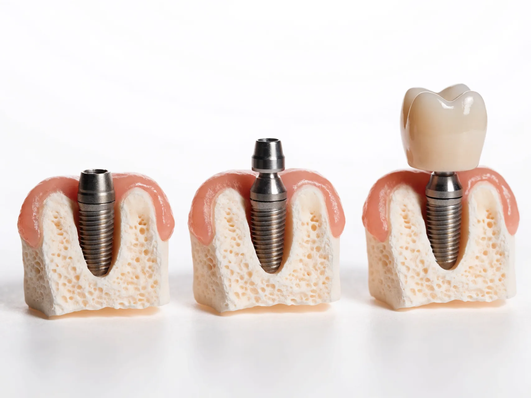
Dental implants after orthodontics are not governed by one waiting period. Braces or aligners may have created a space, redistributed roots, corrected a bite or prepared several teeth for a future restoration, but the end of active tooth movement is only one decision point. An implant is ankylosed to bone and cannot subsequently move like a natural tooth. Its position must therefore be chosen for the planned crown, the neighbouring roots, the gum architecture, long-term cleaning and the retention strategy—not merely for the gap that can be seen in a photograph.
This guide is for adults and families researching an implant after fixed braces, clear aligners, lingual appliances, growth-related treatment or combined orthodontic and restorative care. It also covers congenital absence, trauma, a tooth lost during treatment and a space deliberately opened or maintained. It does not diagnose growth completion, prescribe a retainer, interpret an image or decide that an implant is necessary. Those decisions belong to named clinicians who have examined the patient and accepted responsibility for their part of the plan.
WeCare is not the treating dental provider. Its role is limited to enquiry and referral coordination and, if separately agreed, non-clinical travel logistics only to the extent recorded in writing. Before sharing health information or paying, identify the named legal treatment provider, named orthodontist, named restorative clinician and named implant clinician. Confirm registration, complaint arrangements, record ownership and who will make decisions if the post-orthodontic space or tissue differs from a remote estimate.
The missing-tooth diagnosis comes before the implant
“A gap after braces” is not a diagnosis. The tooth may never have developed, may be impacted, may have been lost after trauma, may have a root or periodontal problem, or may have been removed as part of another treatment. Some apparent gaps are temporary stages in orthodontic space redistribution. Other cases involve a retained primary tooth, an unerupted permanent tooth or a restorative problem that could be treated without extraction. Each pathway has different records, alternatives and sequencing.
Ask the clinicians to name the diagnosis for each site and to explain whether a useful tooth can be retained. AAE guidance on [implants and the preservation of natural teeth](https://www.aae.org/specialty/wp-content/uploads/sites/2/2019/04/Implants_PositionStatement_v2.pdf) cautions against treating extraction and implant placement as automatically preferable to preserving a restorable tooth. If a tooth has uncertain vitality, root damage, resorption, trauma or previous root-canal treatment, obtain a tooth-specific prognosis and appropriate specialist opinion before agreeing to irreversible removal.
For congenital absence, planning should include the whole dentition rather than treating one empty space in isolation. Tooth size, shape, symmetry, remaining primary teeth, other missing teeth, occlusion, bone and facial growth may all affect the choice between space closure, space opening and replacement. The systematic review on [interventions for retained deciduous teeth in severe hypodontia](https://pubmed.ncbi.nlm.nih.gov/29461745/) found a limited evidence base, which is a reason for transparent individual planning rather than a universal extraction rule.
Growth is not proved by finishing braces
Removal of an orthodontic appliance does not itself prove that craniofacial growth and dentoalveolar change have ended. Calendar age, legal adulthood, height, a single image, a statement that growth “looks complete” and eruption of a particular tooth are not interchangeable with an individual growth assessment. Continued vertical facial and alveolar change may alter the relationship between an implant-supported crown and adjacent natural teeth because natural teeth can continue to erupt or adapt while an osseointegrated implant does not follow in the same way.
The ITI review of [dental implants in the growing patient](https://forum-implantologicum.iti.org/feature-article/dental-implants-in-the-growing-patient-3402?languageId=en_US) describes why growth pattern and implant location matter. A systematic review of [implant infraposition and continued craniofacial growth](https://pubmed.ncbi.nlm.nih.gov/32835562/) found a long-term risk of implant-supported crowns becoming infraoccluded relative to neighbouring teeth, while also noting limitations in the available evidence. These sources do not supply an online age or a universal test. They support an individual, documented decision.
If growth is a concern, ask which observations the orthodontic and restorative teams are using, how they will compare records over time, what uncertainty remains and what happens if change continues. Some patients may be better served for now by orthodontic space closure, retention of a useful primary tooth, a removable option, an adhesive bridge or another reversible restoration. “Not yet” should come with a maintenance and review plan rather than pressure to book an arbitrary future date.
Three-dimensional space is more than the visible gap
An implant and crown need coordinated space in several dimensions. The mesiodistal gap seen from the front is only one part. The clinician must consider the distance and angulation of neighbouring roots, buccolingual ridge volume, vertical restorative space, emergence profile, papilla support, contact positions, occlusal clearance and access for cleaning. A crown can appear to fit between teeth while the roots beneath converge into the intended implant path.
Orthodontic records should therefore explain not just that space was opened but what the restorative target was. Ask whether the orthodontist and restorative clinician agreed a proposed crown width and midline, how tooth proportions were assessed and whether the roots were moved into a position compatible with the planned implant. If a lateral incisor is absent, for example, the visible crown space, root divergence, central-incisor symmetry, canine form and gingival levels may pull the plan in different directions. A surgical fixture should not be used to compensate casually for unresolved restorative geometry.
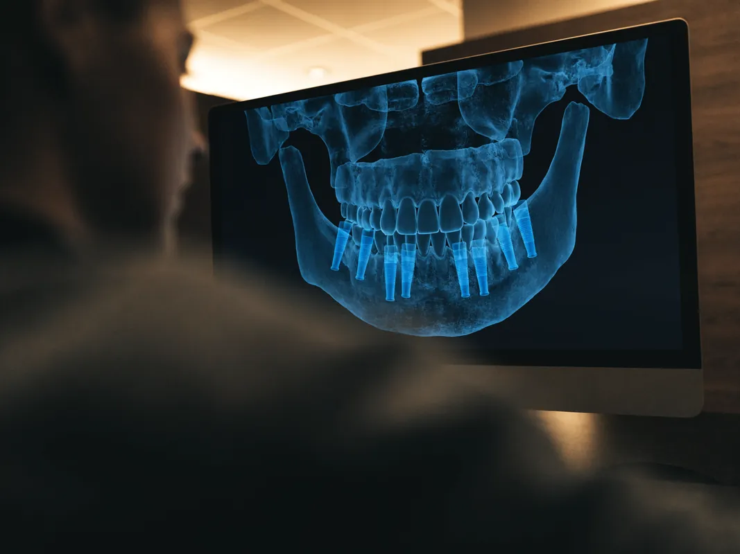
Do not assume that a panoramic image alone can answer these questions. It can contribute useful overview information, yet distortion and two-dimensional projection limit precise spatial interpretation. Intraoral examination, photographs, periodontal assessment, study models or digital scans, appropriate radiographs and—only when justified for the decision—cross-sectional imaging may be combined. Imaging should follow clinical need and radiation-protection principles, not a marketing protocol that promises the same scan to everyone.
Root position must be verified, not inferred from straight crowns
Orthodontic treatment can align the crowns while neighbouring roots remain angled. An attractive finishing photograph cannot show root proximity. The orthodontist should record whether root position and parallelism are suitable for the agreed restorative site, and the implant clinician should independently confirm the surgical anatomy. If the roots are too close or the corridor is unsuitable, options may include additional orthodontic movement, a different restorative design, maintaining the space temporarily or not using an implant.
Root movement also needs periodontal context. Excessive movement outside the supporting envelope may be associated with dehiscence, recession or other tissue concerns in susceptible sites. The systematic review on [gingival recession after orthodontic treatment](https://pubmed.ncbi.nlm.nih.gov/29911278/) found insufficient high-quality evidence to give simple predictions. That uncertainty should lead to a site-specific periodontal examination, not an assumption that all moved teeth have either healthy or damaged supporting tissues.
Ask for dated pre-treatment and completion records where available. These may include photographs, intraoral scans or models, radiographs taken for a justified purpose, periodontal findings and the original orthodontic-restorative objectives. A future clinician should be able to see what changed, why the space exists and whether the final root positions match the intended replacement. Screenshots from a messaging application are not a substitute for original diagnostic files.
Orthodontic retention is part of restorative planning
Teeth can relapse or continue to change after active orthodontic treatment. The British Orthodontic Society's [patient information on orthodontic risks](https://www.bos.org.uk/wp-content/uploads/2023/05/RisksOfOrthodonticTreatment-AdviceSheet-2023.pdf) discusses relapse and the need for retention, while also explaining other matters such as root resorption and periodontal effects. Retention is not merely an administrative phase after the “real” treatment. When a space is being preserved for a restoration, movement can alter contacts, symmetry and the surgical corridor.
The orthodontist should prescribe the retainer type and wear plan for the individual patient. This page does not impose full-time, night-time or lifelong wear, and it does not tell anyone to modify an appliance. Ask how the retainer maintains the implant site, whether it needs a temporary tooth, how it will be adjusted around surgery, who will replace it if it breaks and how the final crown changes the design. A fixed retainer, removable retainer and clear overlay have different practical implications; none should be altered without the responsible clinician.
Retention planning also needs to anticipate access. A retainer that presses on healing tissues, a temporary tooth that transfers unwanted load or an appliance that the patient cannot keep clean may require modification. Conversely, leaving the space uncontrolled while travelling can allow movement that disrupts the planned restoration. The written plan should identify which clinician owns each adjustment and how the patient obtains help away from the orthodontic practice.
Orthodontic finishing records need a formal restorative sign-off
The end of active tooth movement should produce more than a photograph of straight crowns. Before implant surgery, the orthodontic and restorative teams should confirm that the intended space, neighbouring root positions, midline, bite, periodontal condition and retention design correspond to the proposed crown. This is a clinical handover, not proof that every later gate has been met.
The sign-off packet may include the original diagnosis and missing-tooth objective, dated pre-treatment and finishing photographs, scans or models, justified radiographs, root-position assessment, periodontal findings, bite and space measurements, retainer prescription, temporary-tooth design and unresolved issues. Records should distinguish observed findings from predictions. A digital setup can communicate an intended shape but cannot prove tissue stability or available bone.
Ask the restorative clinician to acknowledge receipt and state whether the proposed crown remains feasible. Ask the implant clinician to verify that the surgical corridor and restorative position can be assessed from adequate current information. If either professional wants more orthodontic movement or another examination, clarify who reopens treatment, how retention continues and what cost changes.
The [British Orthodontic Society retainer information](https://bos.org.uk/patients/retainers/) explains that tooth movement after braces is managed with an individually selected retainer and instructions. It does not authorise an implant or prescribe one wear schedule for this page. Its relevance is that retention remains active care while the restorative decision develops.
Retainer failure and space loss need a reversible response
A retainer that breaks, loosens or no longer fits may signal appliance damage, tooth movement, tissue change or interference from a temporary or surgical stage. Do not force it, heat it, cut it or use household adhesive. Contact the responsible orthodontic or dental clinician so the cause and urgency can be assessed. A coordinator cannot determine from a photograph whether a root, crown or implant corridor has changed.
The first response should preserve options. Depending on findings, that may involve repairing or replacing an appliance, adjusting a temporary tooth, taking new records, reopening limited orthodontics, modifying the restorative design or continuing observation. It does not automatically mean that implant placement must proceed quickly before more space is lost.
Document the intended space and root relationship before and after a problem. A visible millimetre change does not alone reveal three-dimensional root position, tissue health or crown feasibility. If movement alters the plan, obtain a revised explanation, alternatives and quotation rather than asking the surgeon to compensate through implant angulation.
Retention responsibility should survive travel and discharge. Ask who provides replacement appliances, how quickly a local review can occur, what the patient pays and how the original orthodontist communicates with the restorative and implant teams. The [NHS braces guidance](https://www.nhs.uk/tests-and-treatments/braces/) confirms that retainers are used after braces and that individual wear advice comes from the orthodontist; it is not an implant timetable.
Do not replace one timetable myth with another
There is no universal interval after braces, aligners or jaw surgery that makes every implant site ready. Readiness can depend on growth, reason for the missing tooth, periodontal condition, root position, stability of the orthodontic result, bone and soft tissue, need for additional movement, type of surgery, medical history, smoking or nicotine exposure, restoration design and ability to maintain aftercare. The calendar is a scheduling tool, not biological proof.
Published work on [adjunctive orthodontic applications in implant dentistry](https://pubmed.ncbi.nlm.nih.gov/24175964/) describes orthodontic site development and space management but found no randomised controlled trials in the reviewed evidence. A systematic review on [treatment of severe hypodontia](https://pubmed.ncbi.nlm.nih.gov/26899287/) similarly reported heterogeneous and generally weak evidence. A responsible clinician can use experience and guidelines while acknowledging where comparative evidence is limited. A website should not convert that uncertainty into a fixed countdown.
Ask what clinical milestone is being awaited. It might be confirmed growth stability, completion of root movement, periodontal stability, healing from another procedure, a stable retainer arrangement or approval of the restorative design. Ask how the milestone will be measured, who will decide and what alternatives remain if it is not achieved. A date should follow the decision criteria, not replace them.
Build one orthodontic-restorative-surgical plan
The Royal College of Surgeons' [standards for dental implant treatment](https://www.rcseng.ac.uk/-/media/Files/RCS/FDS/Publications/Implant-guidelines.pdf) emphasise assessment, planning and the need for multidisciplinary management in complex circumstances including hypodontia and young patients. In a post-orthodontic case, the sequence should be agreed before irreversible treatment: what position and shape should the final tooth have, can orthodontics produce and retain the required space, what tissue is present, where can a fixture be placed, and how will the crown be maintained?
The restorative endpoint should guide the surgical position. A surgeon should not place an implant simply in the centre of available bone if that would force an uncleanable, unaesthetic or functionally compromised crown. Equally, a desired crown position cannot override anatomical safety or tissue limitations. Digital planning, guides and mock-ups may help communication, but they do not turn a plan into a guarantee. Ask which records were aligned, who approved the proposed tooth position and how intraoperative findings could change the plan.
Record the responsibility map. The orthodontist owns tooth movement and retention instructions. The implant clinician owns the surgical diagnosis, consent and procedure. The restorative clinician owns the prosthetic design and delivery. A periodontal, endodontic, paediatric or oral-surgery opinion may be needed for a specific question. If one clinician performs several roles, each decision still needs to be documented. A coordinator cannot assume clinical ownership.
Space closure may be a real alternative
An implant is not the default destination of every orthodontically opened gap. Depending on diagnosis, facial and dental proportions, occlusion, age, growth, tissue and patient preference, space closure may avoid a prosthetic replacement. In other cases, opening or maintaining space for an adhesive bridge, conventional bridge, removable prosthesis, autotransplantation or implant may better fit the overall plan. Each option changes later maintenance and should be explained without presenting one as universally superior.
For selected growing patients and suitable donor teeth, tooth autotransplantation may be discussed by an appropriately experienced multidisciplinary team. The British Orthodontic Society's [information on tooth autotransplantation](https://bos.org.uk/wp-content/uploads/2024/12/TOOTH-AUTOTRANSPLANTATION-2024.pdf) explains its role and limitations. It is not available or appropriate for everyone, and this page does not recommend it. Its relevance is that irreversible implant treatment should not be presented before reasonable tooth-based and reversible alternatives have been considered.
When comparing options, ask about effects on healthy neighbouring teeth, need for future replacement, maintenance access, tissue appearance, retention, possibility of revision, local provider availability and what happens as the dentition changes. “Do nothing for now” can be an informed choice if risks, monitoring and temporary appearance or function are understood.
Periodontal health and soft-tissue architecture
Straight teeth do not prove periodontal health. Before implant surgery, the clinician should examine plaque control, bleeding, pocketing, recession, mucogingival conditions, active infection, tooth mobility and the patient's ability to clean the site. Previous orthodontic appliances may have made cleaning difficult; conversely, alignment may have improved access. Neither direction should be assumed. Active disease should be diagnosed and addressed before an elective implant decision.
The appearance of the future crown depends partly on soft-tissue and bone relationships that were shaped before and during orthodontics. Papilla height is influenced by neighbouring teeth and their supporting tissues. A missing tooth after trauma or long-standing absence may have a different ridge contour from a recently created space. Ask whether a periodontal or restorative assessment is needed before further tooth movement ends, because some tissue decisions are easier to coordinate before the orthodontic appliances are removed.
A promise that a graft will “fix” every contour is unsafe. The need, type, source, timing and limitations of any graft should be decided after examination and appropriate imaging. Ask what defect is being treated, which alternatives exist, what material is proposed, what traceability information will be supplied and how a change in findings would affect the procedure and quotation. Consent should distinguish an expected component from a contingency.

Implant planning still needs a complete dental and medical assessment
Post-orthodontic status is only one part of implant assessment. The named clinician should examine the proposed site, remaining teeth, gums, bite, oral hygiene, parafunction, mucosa and restoration space. The history should cover the reason for tooth loss, previous orthodontic and surgical treatment, smoking or vaping, allergies, previous healing, relevant medical conditions and all current prescribed and non-prescribed medicines and supplements.
Do not stop, delay or change a medicine because of a website or travel date. If a dental procedure raises a specific question about anticoagulants, antiresorptive therapy, diabetes management, immune treatment, pregnancy, sedation or another issue, the dental clinician should seek focused input from the responsible medical professional with the patient's permission. A generic “medical clearance” label is less useful than a precise question linked to the proposed intervention.
Imaging should answer a defined diagnostic or planning question. Ask why each image is needed, who reports it and how it changes the options. Cross-sectional imaging may be justified when two-dimensional information is insufficient for anatomical or surgical planning, but it is not a universal post-braces ritual. A remote panoramic image cannot confirm root clearance, tissue quality, periodontal health or final implant suitability by itself.
Separate surgical readiness from restorative readiness
An implant can be surgically placeable while the definitive crown design remains unresolved. That is not an acceptable reason to improvise later. Before surgery, the restorative clinician should define the proposed tooth position, contour, contacts, occlusion, material considerations, retention method, cleanability and likely maintenance route. If the implant angle or depth constrains those choices, the patient should understand the implications before placement.
The converse also occurs: an ideal digital crown can be drawn where anatomy does not permit a safe or maintainable fixture. The surgical and restorative teams need to reconcile those constraints. A diagnostic wax-up, digital setup or trial smile can illustrate a target, but it remains a planning aid. It cannot promise papilla fill, colour match, tissue stability or a specific appearance.
If veneers, bonding or crowns on neighbouring teeth are being considered, sequence them deliberately. Tooth whitening should be planned before definitive shade selection when clinically appropriate, because ceramic restorations do not whiten like natural teeth. Additive bonding may offer a reversible way to adjust tooth proportions. Preparing healthy teeth only to make an implant crown appear symmetrical requires a separate consent discussion about lost tooth structure and alternatives.
Provisional and final restorations are different decisions
A temporary tooth used during orthodontics, a retainer pontic, an adhesive temporary, a provisional implant crown and a definitive crown have different purposes and loading conditions. “You will have a tooth throughout” is too vague. Ask exactly what will be present at each stage, whether it is removable or fixed, what it is supported by, whether it contacts the bite, how it is cleaned and what happens if it loosens or fractures.
Immediate provisionalisation is not automatically available after orthodontics. The decision can depend on site anatomy, primary stability, tissue, implant position, occlusion, parafunction and the clinician's case-specific protocol. A temporary appearance should not be confused with biological integration or a final restoration. The clinician must explain what findings allow or rule out each option and what alternative temporary arrangement will be used if the planned one is not appropriate.
Final restoration should follow clinical evidence of readiness rather than a universal number of weeks or months. Ask what findings are reviewed, whether soft-tissue shaping is needed, how the bite is assessed and which records form the baseline. If further orthodontic movement or retainer changes are anticipated, the final contacts and appliance compatibility need joint approval.
Consent is a continuing process
The General Dental Council's [Principle 3 on valid consent](https://standards.gdc-uk.org/pages/principle3/principle3) is written for UK registrants, but it provides a useful patient benchmark: options, risks, benefits, likely prognosis, possible costs and the consequences of no treatment should be discussed, and consent remains valid only while the patient understands and agrees. An overseas provider is governed by its own jurisdiction; citing GDC standards does not prove that provider's regulation.
Post-orthodontic consent should identify uncertainty about growth, relapse, root position, tissue appearance and any need for additional orthodontics or grafting. It should cover implant and restorative risks, effects on adjacent structures, temporary arrangements, alternatives, maintenance and the possibility that examination changes the plan. The patient can defer or refuse treatment. A deposit, flight or completed orthodontic phase does not remove that choice.
If the plan changes after examination, ask for the new diagnosis, reasons, alternatives, risks, itemised cost and schedule implications before consenting. Do not accept a same-day sales decision for an unanticipated extraction, graft, extra implant or alteration of healthy teeth unless delay would itself create an urgent clinical problem and the responsible clinician explains why.
Records should connect the orthodontic and implant phases
The GDC's [Principle 4 on patient information](https://standards.gdc-uk.org/pages/principle4/principle4) describes accurate records, including an updated medical history and, where available, radiographs, consent documents, photographs, prescriptions and referral correspondence. Use this as a practical checklist even when care occurs elsewhere. Ask who holds the complete record and how original files will be transferred securely.
Useful pre-treatment records may include the original diagnosis, orthodontic objectives, missing-tooth and trauma history, dated photographs, scans or study models, relevant radiographs, root-position assessment, retention prescription, periodontal findings, correspondence between clinicians and the intended restorative design. After implant treatment, request procedure notes, implant and component identifiers, graft or membrane identifiers where used, anaesthetic and medicine records, images, restoration laboratory information, occlusal and periodontal baseline, aftercare instructions and direct clinical contacts.
The record should be understandable to a local dentist who did not sell or perform the treatment. A branded implant passport without diagnosis, procedure detail or restoration information is incomplete. Keep copies in accessible formats and give permission for necessary transfer between the named clinicians.
Demand an itemised quotation and change-control rules
A quotation should separate assessment, imaging, orthodontic review or adjustment, periodontal care, implant surgery, components, possible grafting, temporary replacement, definitive crown, laboratory work, medicines, protective appliance, reviews and record delivery. Non-clinical travel services should be listed separately. Ask whether tax, revisions, remakes, additional visits, local care and management of complications are included, excluded or undecided.
Do not treat the quoted number as a diagnosis. A preliminary estimate can change after examination, but the provider should explain the reason and obtain new consent before non-urgent additional treatment. Ask how deposits, cancellation, postponement, refunds, incomplete treatment and disputes are handled. Any warranty is a commercial term with conditions and exclusions; it does not guarantee a biological or aesthetic result and does not replace accessible clinical aftercare.
Compare like with like. One proposal may include a provisional restoration and final crown while another quotes only fixture placement. One may assume no graft and another may price a contingency. Ask for product identities and traceability, but do not use a brand name as a substitute for clinician competence, restoration design or maintenance planning.
Travel must follow the clinical sequence
The GDC's [guidance on going abroad for dental treatment](https://www.gdc-uk.org/standards-guidance/information-for-patients-public/going-abroad-for-dental-treatment) advises patients to verify qualifications and regulation, understand the treatment plan and costs, allow time for recovery, and know the aftercare and complaint arrangements. The NHS [treatment-abroad checklist](https://www.nhs.uk/using-the-nhs/healthcare-abroad/going-abroad-for-treatment/treatment-abroad-checklist/) also prompts questions about risks, complications, records, insurance and follow-up.
Travel dates should be built after the orthodontic, surgical and restorative teams define decision points. Do not buy inflexible flights on the assumption that appliance removal, surgery, provisionalisation or final restoration will occur on advertised dates. Ask how long is needed locally for assessment and review, who can change fitness-to-fly advice, and what happens if swelling, infection, pain, altered sensation, retainer breakage or an unexpected procedure delays travel.
Check travel insurance in writing. Routine policies may exclude planned treatment, consequences of elective care, pre-existing conditions or extra accommodation and flights. A coordinator's reassurance is not the policy. Local treatment and no travel must remain genuine alternatives, particularly when orthodontic follow-up is still active or the patient lacks a nearby clinician willing to provide maintenance.
Local aftercare and retention handover
Implant maintenance and orthodontic retention continue after travel. Before treatment, identify a local dentist and, where needed, the original orthodontist. Ask whether they are willing to review the planned restoration, adjust the retainer, monitor tissues and communicate with the treating clinic. Do not assume that an uninvolved local professional will repair another provider's work, hold compatible components or accept clinical responsibility without records and examination.
The handover should say how to clean the restoration, how the retainer is used, what symptoms are expected, what changes need routine review and what requires urgent assessment. It should identify the destination clinician's contact route and the local pathway. Review intervals should be based on individual need rather than one universal schedule. Baseline probing and radiographic information, when clinically appropriate, helps later clinicians recognise change.
If the retainer no longer fits, do not force it over a surgical or restored site and do not cut it at home. Contact the responsible orthodontist or dentist promptly. If adjacent teeth begin to move, early assessment may preserve options. If a temporary tooth loosens, keep it if safe to do so and seek instructions rather than using household adhesive.
Red flags in a post-orthodontic implant proposal
Pause when a seller says the end of braces automatically proves readiness; gives the same wait to every patient; ignores age or growth questions; measures only the visible gap; cannot show how neighbouring roots were assessed; proposes extracting a potentially restorable tooth without a prognosis; or books surgery before a restorative design exists.
Other warnings include no named clinician, no direct clinical consultation, no periodontal assessment, routine cross-sectional imaging without justification, a promise that grafting will guarantee aesthetics, a fixed final crown date before examination, pressure to alter several healthy teeth, no retainer plan, no product traceability, no itemised quotation, a guarantee presented as an outcome, and no local aftercare pathway.
Be cautious if clinicians do not communicate with one another or if each says another person owns the decision. A patient should not have to mediate conflicting instructions about space, roots, retention, surgery and the final crown. Written multidisciplinary agreement is especially important in congenital absence, trauma, continued growth, jaw surgery, complex tissue defects or several missing teeth.
Urgent and emergency boundaries
Seek urgent local dental assessment for worsening or persistent swelling, increasing pain, discharge or bad taste, fever, uncontrolled bleeding, a loose implant or restoration, new or worsening altered sensation, inability to clean or eat as instructed, or a retainer or appliance causing injury. A remote coordinator cannot examine infection, nerve symptoms, tissue breakdown or occlusal instability.
For people in England, the NHS explains how to obtain [urgent or emergency dental care](https://www.nhs.uk/nhs-services/dentists/how-to-find-an-nhs-dentist-in-an-emergency/). Severe swelling affecting breathing or swallowing, uncontrolled heavy bleeding, major facial trauma or serious systemic illness requires emergency medical help. Use the relevant local emergency route where you are. These boundaries are not a prediction that complications will occur; they are part of responsible planning.
Questions for the combined clinical team
- What is the diagnosis and why is the tooth absent or proposed for removal?
- Can any useful tooth be preserved, and has the appropriate specialist assessed it?
- Is growth relevant, what evidence is being reviewed and what uncertainty remains?
- What final crown position and dimensions guided the orthodontic space target?
- Are the neighbouring roots positioned for the proposed implant corridor?
- What periodontal and soft-tissue findings affect the plan?
- Which options were compared: space closure, temporary replacement, bridge, autotransplantation, implant or no treatment?
- What retention appliance is prescribed before, during and after implant care?
- Who adjusts or replaces the retainer around surgery and restoration?
- Which clinician owns orthodontics, surgery, restoration and long-term maintenance?
- What imaging is justified, and how could its findings change the proposal?
- What is provisional, what is definitive and what findings separate the stages?
- What materials and component records will be supplied?
- What is itemised, contingent, excluded or separately payable?
- What local aftercare is arranged and what urgent route applies after travel?
Post-orthodontic implant planning checklist
Diagnosis and preservation
- Record why each tooth is missing or compromised.
- Obtain a tooth-specific prognosis before extraction.
- Compare preservation, space closure, bridge, temporary and no-treatment options.
Growth, space and roots
- Document how growth relevance was assessed without relying on age alone.
- Define the crown-led target in three dimensions.
- Verify root position and the supporting periodontal tissues.
Retention and sequence
- Keep the orthodontist's individual retention prescription.
- Plan retainer adjustments around surgery and restoration.
- Separate provisional appearance, integration assessment and definitive restoration.
Consent, quote and records
- Identify the legal provider and every responsible clinician.
- Obtain the risks, alternatives, uncertainties and change-control process in writing.
- Request an itemised clinical quotation and separate travel terms.
- Secure original orthodontic, surgical, component and restorative records.
Travel and continuity
- Build travel around clinical decision points, not an advertised countdown.
- Confirm insurance, flexible arrangements and fitness-to-fly advice.
- Identify local orthodontic, dental, urgent and emergency routes before leaving.
Sources and evidence limits
- [Royal College of Surgeons, Guidelines for Selecting Appropriate Patients to Receive Treatment with Dental Implants](https://www.rcseng.ac.uk/-/media/Files/RCS/FDS/Publications/Implant-guidelines.pdf), accessed 29 August 2026. It supports comprehensive assessment, consent, restorative planning and multidisciplinary management, including young patients and hypodontia; it does not create an individual treatment decision.
- [ITI, Dental implants in the growing patient](https://forum-implantologicum.iti.org/feature-article/dental-implants-in-the-growing-patient-3402?languageId=en_US), accessed 29 August 2026. It explains growth-related principles and implant-location considerations without providing a universal age threshold.
- Bohner and colleagues, [systematic review of implant infraposition and craniofacial growth](https://pubmed.ncbi.nlm.nih.gov/32835562/), 2020. It supports disclosure of long-term relative-position change and evidence limitations.
- Uribe and colleagues, [systematic review of adjunctive orthodontic applications in implant dentistry](https://pubmed.ncbi.nlm.nih.gov/24175964/), 2013. It covers orthodontic site development and space management while reporting the absence of randomised controlled trials in the reviewed evidence.
- Filius and colleagues, [systematic review of oral rehabilitation in severe hypodontia](https://pubmed.ncbi.nlm.nih.gov/26899287/), 2016, and Laverty and colleagues, [systematic review of retained deciduous teeth](https://pubmed.ncbi.nlm.nih.gov/29461745/), 2018. They demonstrate heterogeneous approaches and limited evidence rather than a standard replacement sequence.
- Tepedino and colleagues, [systematic review of gingival recession after orthodontic treatment](https://pubmed.ncbi.nlm.nih.gov/29911278/), 2018. It supports cautious, site-specific periodontal assessment rather than a universal prediction.
- [British Orthodontic Society, Risks of Orthodontic Treatment](https://www.bos.org.uk/wp-content/uploads/2023/05/RisksOfOrthodonticTreatment-AdviceSheet-2023.pdf), 2023, and [Tooth Autotransplantation](https://bos.org.uk/wp-content/uploads/2024/12/TOOTH-AUTOTRANSPLANTATION-2024.pdf), 2024. These patient resources support retention, risk discussion and awareness of a selected tooth-based alternative.
- [General Dental Council Principle 3](https://standards.gdc-uk.org/pages/principle3/principle3), [Principle 4](https://standards.gdc-uk.org/pages/principle4/principle4) and [going-abroad guidance](https://www.gdc-uk.org/standards-guidance/information-for-patients-public/going-abroad-for-dental-treatment), accessed 29 August 2026. They are UK patient-facing benchmarks for consent, records and provider checks, not evidence that an overseas provider is UK-regulated.
- [NHS treatment-abroad checklist](https://www.nhs.uk/using-the-nhs/healthcare-abroad/going-abroad-for-treatment/treatment-abroad-checklist/) and [urgent dental guidance](https://www.nhs.uk/nhs-services/dentists/how-to-find-an-nhs-dentist-in-an-emergency/), accessed 29 August 2026. They support travel, aftercare and escalation planning.
For broader context, see the dental implant service guide, young-adult implant planning guide, treatment-stage guide and returning-home aftercare guide. These links add planning context; none replaces examination by the responsible clinicians.