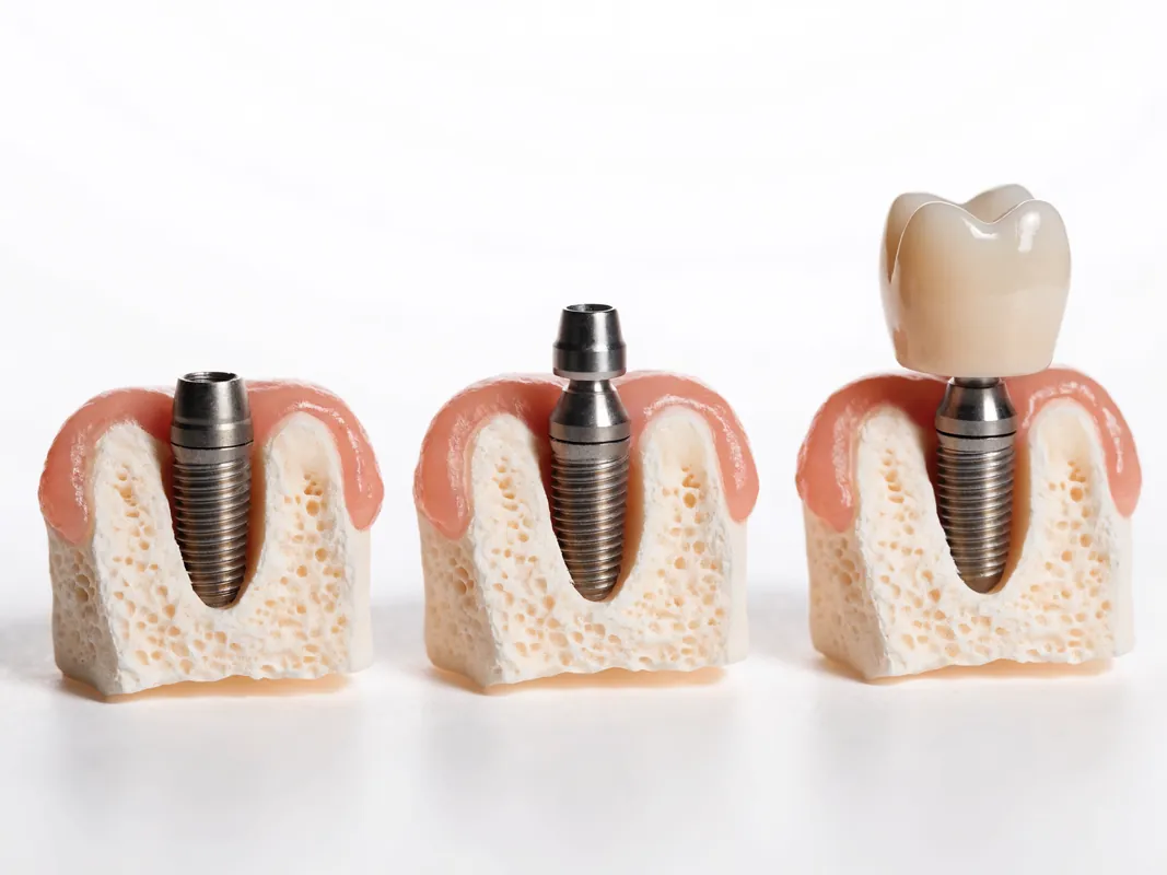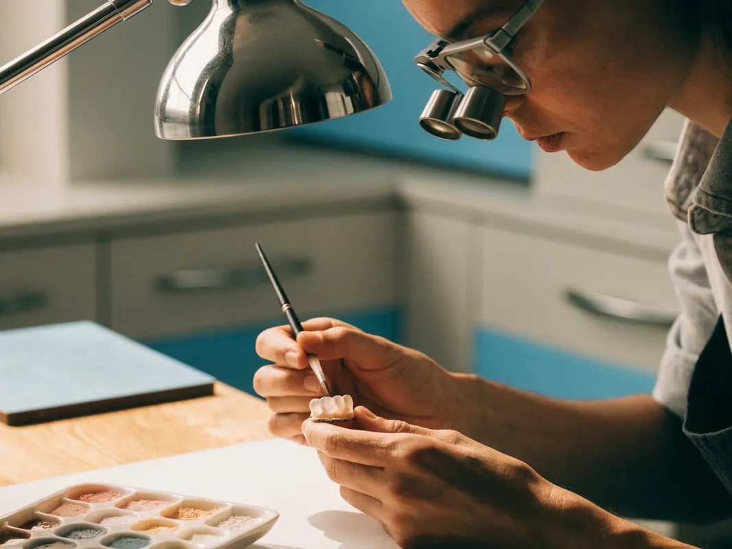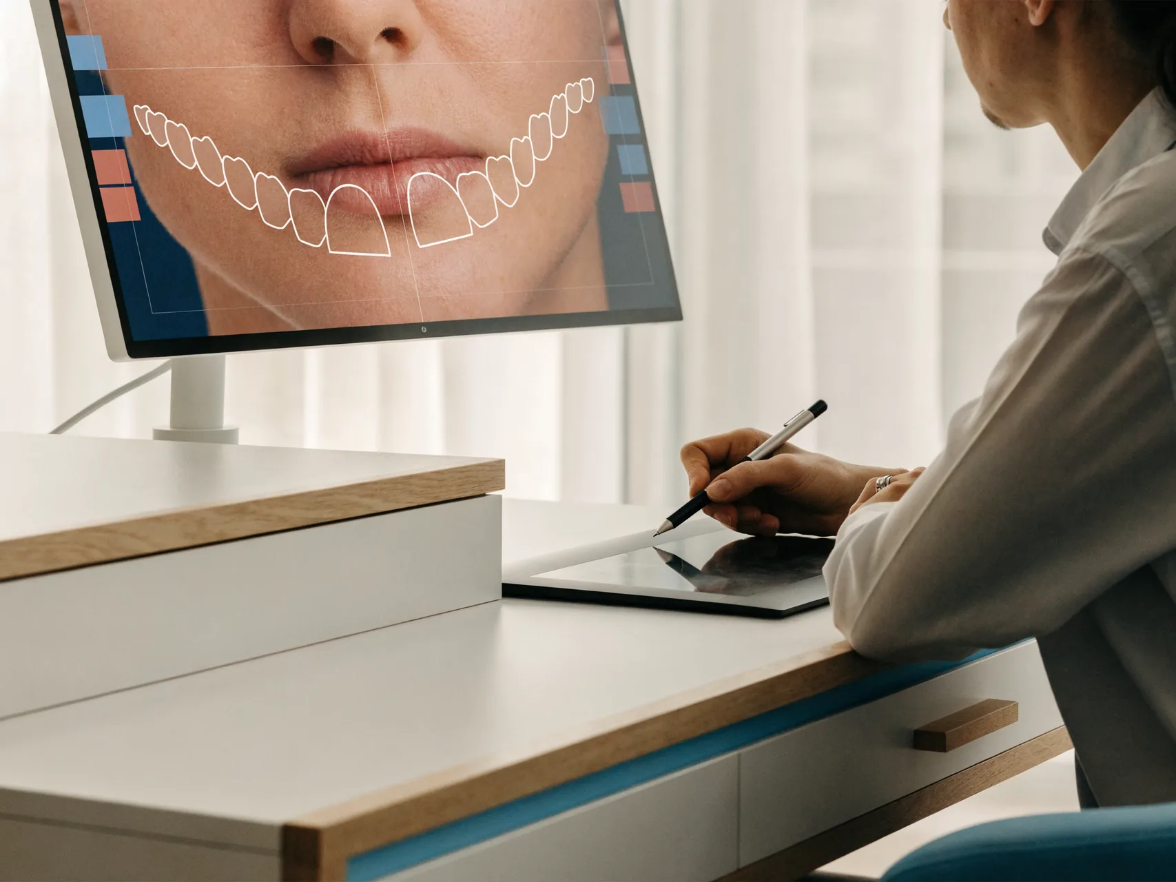
Digital smile design is a planning tool, not a treatment. It can bring photographs, scans, measurements and a patient's preferences into one discussion about possible changes to tooth colour, shape, position and the smile line. It does not diagnose disease, choose a material by itself or make an irreversible procedure necessary.
A digital preview may not reproduce texture, translucency, lip movement, gum response or the way a restoration will look in changing light. A preview is not a guarantee of the final result. Its value is in making questions and trade-offs visible before consent, not in promising a perfect smile.
This guide is for people comparing digital smile design in Turkey, a local planning consultation or a cross-border restorative proposal. It does not prescribe a scanner, camera, software, algorithm, tooth shape, material, procedure, appointment count, price or travel schedule. The responsible clinicians must decide which records are justified and how any design relates to oral health, function and tissue preservation.
The [GDC consent standard](https://standards.gdc-uk.org/pages/principle3/principle3) explains that consent is a continuing discussion about options, risks, potential benefits and costs, not a signature alone. The [GDC patient-record standard](https://standards.gdc-uk.org/pages/principle4/principle4) includes photographs, models, recordings, laboratory prescriptions and conformity information where available. UK data questions can also be informed by the [ICO health-data guidance](https://ico.org.uk/for-organisations/uk-gdpr-guidance-and-resources/lawful-basis/special-category-data/what-is-special-category-data/). These sources provide a question framework; they do not regulate or endorse a Turkish provider.
Digital smile design is not one standard product
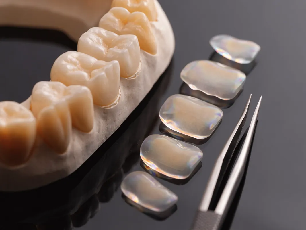
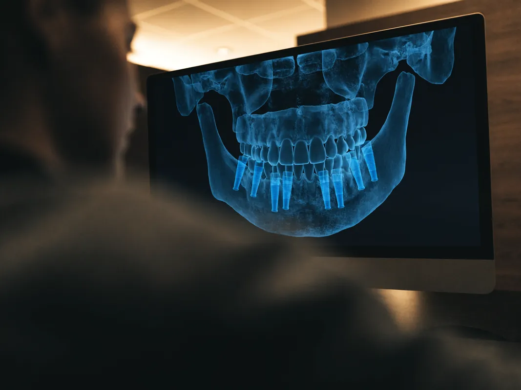
The phrase can describe very different workflows: a simple photograph with lines drawn over it, a two-dimensional simulation, an intraoral scan, a facial scan, a digital wax-up, a physical printed model, an additive trial smile, provisional restorations or a combination of these. A provider should state exactly what is included and what clinical decision each record supports.
A branded label or software screenshot does not establish diagnostic quality. The useful evidence is the named clinical owner, calibrated source records, current file versions, tooth-by-tooth objectives, laboratory prescription, mock-up or provisional criteria, consent gates and final treatment record. If the design is only a communication image, it should be labelled that way.
Do not pay for an undefined design package. Ask which outputs you receive, whether the files can be opened outside the provider's system, who owns or controls them, and what happens if you choose no treatment.
Who may find planning useful?
Planning may be useful for adults considering visible changes involving several teeth, orthodontics, bonding, whitening, veneers, crowns, implants or gum treatment. It can help a patient explain preferences, compare conservative alternatives and understand which parts of an image are simulation rather than anatomy.
Not everyone needs digital smile design. A person seeking a routine examination, hygiene care, a small filling or relief from pain usually needs diagnosis first. Someone with active decay, gum disease, an unstable bite, tooth wear, missing teeth or unexplained symptoms may need those issues assessed before cosmetic options are modelled. Eligibility for any later treatment depends on oral health, tooth structure, bite, medical history and the clinician's examination.
Bring a list of medicines, relevant medical conditions, past dental treatment, concerns about grinding or jaw symptoms, and any existing radiographs or scans. Reference photographs can communicate taste, but another person's teeth cannot be copied safely onto a different face and bite.
Define the decision before collecting records
Every scan, photograph and simulation should answer a stated question. Examples include whether an additive change could address a chipped edge, how proposed tooth length may affect speech, whether a space can be closed without creating excessive width, how natural teeth and restorations relate in colour, or whether a provisional phase is needed before definitive work.
Write the question first. Then identify the least intrusive record capable of informing it. A portrait photograph may show tooth display and facial context. An intraoral photograph may show colour, surface and restorations more clearly. A scan can record geometry but not pulpal health, material strength or every subgingival finding. Imaging involving radiation requires a separate clinical justification; a prettier simulation is not a reason to expose a patient.
Record what will not be decided by the design. A visual plan cannot determine whether decay is present under a restoration, whether a tooth is vital, whether gum disease is stable, whether an implant site has adequate anatomy, or whether a requested material is suitable. Those questions belong to clinical assessment.
Name every clinical and technical owner
Ask who examines and diagnoses, who creates the design, who approves tooth movement or preparation, who communicates with the laboratory, who performs each procedure and who remains responsible after treatment. Verify current professional registration and do not infer specialist status from a job title or social-media biography.
A designer, technician, coordinator and treating dentist can contribute different expertise. Their roles should not be blurred. A technician may interpret a prescription and manufacture a model or restoration, but the named clinician must own diagnosis, consent and clinical decisions. A coordinator may arrange files and appointments, but should not convert a provisional image into treatment advice.
If the team or laboratory changes, request an updated plan and explain how responsibility, file transfer, confidentiality and cost change. The patient should know who can answer a clinical question directly.
Assessment comes before the image
A sound assessment records the reason for consultation and examines teeth, gums, bite, smile dynamics and existing restorations. Photographs may document the face and teeth. An intraoral scan or conventional impression may be useful when a physical or digital model is needed. Imaging should have a clinical justification; a cosmetic simulation alone is not a reason for unnecessary radiation.
Ask who performs the examination, who creates the design and who would carry out any proposed treatment. Verify professional registration and relevant experience directly with the regulator and named provider. Software, cameras, scanners and mock-up materials differ, so the provider should confirm in writing which records and planning stages apply to your case. A generic website list is not proof of local inventory.
Remote images may support an initial conversation but are not a final diagnosis. Use the secure method specified by the provider for health information, and ask how files will be stored, shared with laboratories or other clinicians, retained and deleted.
Build a record set that matches the question
A planning record set may include facial photographs at rest and smiling, intraoral photographs, video of speech and movement, a digital or conventional impression, a bite record, shade information and existing clinical images. Not every patient needs every record. Each item should have a purpose, date, owner and quality check.
Photographs need consistent orientation, focus, exposure and colour handling if they will be compared. A phone portrait can communicate preference but may distort proportion. A retracted intraoral view gives different information from a social smile. Video may show dynamic tooth display and speech but does not diagnose tissue health.
An intraoral scan records visible surface geometry. It can support models, measurements and laboratory communication, but blood, saliva, reflective surfaces, inaccessible margins, movement and scanning technique can affect capture. The scan does not reveal the internal condition of a tooth. Ask which areas were incomplete and whether conventional records are still needed.
A facial scan or merged dataset adds another alignment problem: files must be registered accurately. A persuasive three-dimensional image can still be wrong if captures were taken at different times, jaw positions or expressions. Record the merge method and verify key landmarks clinically.
Separate photographs, scans, models and imaging
These words are often mixed in marketing, but they carry different information and risk:
- a photograph records a two-dimensional visual appearance;
- an intraoral scan records surface geometry;
- a physical or digital model represents selected geometry;
- a facial scan represents external facial surfaces;
- a radiograph or CBCT image uses ionising radiation for a justified clinical question;
- a simulation changes an image or model to display a proposal.
One cannot automatically substitute for another. A scan is not an X-ray. A simulation is not a diagnosis. A model cannot show every soft-tissue response. Imaging should be selected by the responsible clinician, not added because a package is called fully digital.
Ask for the original clinical files and the derived planning files to be distinguished. That separation makes later review, correction and consent more reliable.
What a useful design discussion covers
A useful plan separates preference from clinical necessity. It may discuss tooth display at rest and in speech, midline, proportions, colour, incisal edges, gum levels and the relationship between upper and lower teeth. These are design observations, not a prescription.
A screen rendering is usually two-dimensional even when based on a scan. A physical trial smile or temporary mock-up may add information about speech, lip support and appearance, but it may feel thicker than a final restoration and may not be possible without preparation in every case. Ask whether the mock-up changes any tooth structure and whether it can be removed without damage before agreeing.
The patient should be able to request a calmer, less uniform or more natural direction. Symmetry and very bright shades are preferences, not clinical standards. The clinician should explain where anatomy, function or material thickness limits the requested look.
Two-dimensional previews have predictable blind spots
A photograph-based preview can explore tooth outline, visible length, colour direction and broad symmetry in one captured expression. It cannot reliably show the back of teeth, contact areas, thickness, bite, jaw movement, tongue space, cleanability, preparation depth, internal tooth condition or material behaviour in every light.
Perspective matters. Camera distance, lens, head position, lip movement and image crop can alter perceived proportion. A design drawn on a wide-angle phone image may not transfer accurately to the mouth. Ask whether the photograph was standardised and which measurements are true clinical measurements rather than screen pixels.
Do not let a dramatic slider or instant before-and-after image bypass alternatives. The preview should carry a clear label: illustrative proposal, subject to examination, functional assessment, material limits and renewed consent.
Three-dimensional design still needs clinical verification
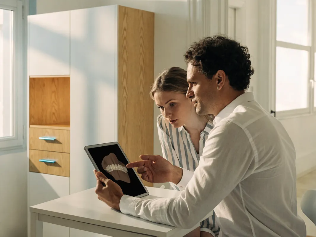
A digital wax-up can model surface form and proposed additions in three dimensions. It may support measurements, a printed model, a guide, a mock-up or laboratory communication. It still depends on scan completeness, bite registration, alignment, design parameters and the assumptions entered by a human or algorithm.
Check proposed thickness, contacts, emergence, contours, connector areas where relevant, opposing clearance and cleaning access on the actual case. A model can appear smooth while producing an overcontoured, difficult-to-clean or functionally unsuitable restoration. The clinician and technician should document how design limits were tested.
Version control matters. Name and date the diagnostic scan, design revision, patient-reviewed version, clinical prescription and manufacturing file. If the mouth changes after provisional treatment, orthodontics, gum healing or tooth preparation, the old design may no longer represent the current anatomy.
Dynamic smile and speech assessment
A smile changes through speech, laughter, head movement and different lip positions. Static symmetry is only one part of the decision. Proposed incisal length, tooth position and contour may influence lip contact, speech sounds, tongue space and the way teeth meet.
Use natural conversation, relevant sounds and ordinary expressions rather than one exaggerated smile. If a mock-up or provisional is available, the patient should have a genuine chance to speak, view it at normal distance and assess comfort and appearance. Record concerns instead of dismissing them because the screen design looked correct.
Jaw symptoms, grinding, wear and an unstable bite require their own assessment. Digital smile design cannot prove that a new contour will be tolerated. A functional concern can outweigh visual preference.
Mock-up is a decision aid
An additive intraoral mock-up can translate part of a design into the mouth without representing the final material. It may help assess visible length, proportion, speech, lip support and general contour. It can feel bulkier than a definitive restoration and may not be feasible without alteration in every case.
Before placement, confirm whether the mock-up is fully reversible, whether any tooth or restoration will be etched, roughened, adjusted or prepared, and how it will be removed. Photograph and document the version reviewed. The patient must be able to request changes or stop without being pressured into drilling.
A mock-up cannot prove final shade, translucency, bond, gum response, strength or lifespan. Treat approval as feedback on a trial, not consent to every later procedure.
Provisional restorations are another clinical gate
Where treatment requires preparation or staged restorative work, provisional restorations may test function, contour, cleanability and appearance while protecting teeth. They are not merely a waiting-room copy of the final design. Record material, fit, contacts, bite, symptoms, tissue response and patient feedback.
Changes learned from provisionals should be transferred deliberately to the definitive prescription. Ask whether a new scan or impression is taken, how approved contours are communicated, and what remains open to change. If symptoms or tissue problems appear, clinical reassessment takes priority over the manufacturing schedule.
The patient should understand what happens if they do not approve the provisional stage, including adjustment, remake, delay, additional cost and the consequences of stopping after irreversible preparation.
Limitations to understand
Digital planning cannot predict biological healing, gum movement, colour stability, fracture, wear or long-term maintenance. It cannot show the exact optical behaviour of a future restoration on every screen. A model may hide decay, cracks, old cement, weak tooth structure or bite forces that only an examination reveals.
A design may also make treatment appear simpler than it is. Moving teeth in software is not orthodontic treatment. Adding a virtual tooth is not implant planning. Changing the outline of a tooth does not show how much healthy tissue would be removed for a veneer or crown. Each proposed procedure needs its own diagnosis, consent, risks, alternatives and fee.
Do not treat approval of an image as consent to drilling. Consent should name the actual procedure and remain revocable before treatment begins.
AI and automation do not own the decision
Software may automate tooth segmentation, landmark detection, proposal generation, library selection or image alteration. An automated output can be useful for exploration, but it inherits training, input and configuration limits. It may favour standardised shapes, miss pathology, misread margins or create a persuasive image that has no safe clinical route.
Ask whether automation or generative tools were used, which parts a clinician reviewed, how errors can be corrected and whether patient data was used beyond the individual case. A provider should not call an output personalised merely because a name was attached to it.
The named clinician must remain accountable for clinical relevance and consent. The patient should be able to reject an algorithmic proposal, compare a human-designed or no-design route, and understand that an AI score is not a diagnosis or outcome prediction.
Calibration and repeatability
Shade and proportion records are useful only when their limitations are understood. Colour varies with lighting, camera settings, display calibration, tooth hydration and surrounding colours. A scan can vary with technique and software processing. Facial posture and expression can change between captures.
Record the capture conditions needed for comparison. Use physical shade references and clinical judgement where colour decisions matter. Keep source files so an edited preview can be compared with the unaltered record. Repeating a capture can test whether a difference is real or a recording artefact.
Do not infer precision from many decimal places or a realistic render. Measurement uncertainty, biological variability and manufacturing tolerance should be explained in terms the patient can use.
Maintain a clear version history
Each design iteration should have a date, author, source records and purpose. Mark which version is illustrative, which is prescribed for a mock-up, which the patient reviewed, which the clinician approved, which the laboratory received and which was manufactured.
Do not overwrite an earlier version without preserving the decision trail. If tooth position, gum contour, preparation, implant components or provisional findings change, identify which dependent files are obsolete. The final clinical record should show what was actually delivered, not only the initial sales rendering.
Version history supports complaint handling, laboratory remakes and local handover. It does not prove that the design was clinically appropriate, but it prevents avoidable confusion about which proposal governed each stage.
Conservative alternatives
Clarify the underlying concern before selecting treatment. Whitening may address colour in natural teeth. Orthodontics may change position without covering teeth. Additive bonding may adjust selected edges or spaces with less removal of tooth structure, although it has maintenance limits. Contouring may suit very small changes. Gum treatment requires periodontal assessment. Sometimes photography, hygiene care or no treatment is the most proportionate option.
Veneers and crowns are not interchangeable planning labels. A crown generally covers more tooth structure and is usually considered for restorative reasons; a veneer is also irreversible when tooth preparation is required. Ask how much healthy tooth structure each option would remove and whether an additive or no-treatment path is reasonable.
Use a tooth-preservation ladder
For each proposed tooth, ask the team to compare reasonable options from least to more invasive where clinically appropriate:
- no treatment or monitoring;
- hygiene, stain removal or preventive care;
- whitening of suitable natural teeth;
- orthodontic alignment or space management;
- minor contouring within safe limits;
- additive composite repair or reshaping;
- repair of an existing restoration;
- a veneer or partial restoration when justified;
- a crown only when structural or restorative findings support greater coverage.
This is not a universal sequence. A broken, heavily restored or diseased tooth may need another route. The purpose is to stop a visual simulation from making irreversible preparation appear inevitable. Request a tooth-by-tooth reason for every rejected alternative.
The design should show proposed additions and reductions separately. A front-view image can hide how much material must be removed from a protruding tooth to fit the illustrated outline.
Gum health and contour are not graphic elements
Software can redraw a gum line instantly. Real gingival tissue has biology, thickness, attachment, inflammation, recession risk and healing behaviour. A digital proposal cannot authorise gum surgery or predict a stable contour.
Periodontal assessment comes before elective contour change. Ask whether asymmetry is caused by inflammation, recession, altered eruption, tooth position, restoration contour or natural variation. Compare disease control, no treatment, orthodontics, restorative design and periodontal procedures where relevant.
If tissue treatment is proposed, name the clinician, diagnosis, sites, intended procedure, risks, healing review and how later scans or designs will be updated. Do not manufacture definitive restorations from a pre-treatment contour without a justified plan for change.
Colour and material need a separate workflow
A bright screen does not represent a ceramic, composite or natural tooth in every light. Natural teeth, existing restorations, substrate colour, material, thickness, translucency, surface texture, cement and laboratory characterisation interact. A generic white shade in a preview is not a material prescription.
Map natural teeth and restorations first. If whitening is suitable, sequence it before final shade decisions when appropriate and reassess whether restorations are still wanted. Read the professional teeth whitening service and veneers and whitening guide for those separate gates.
Request the exact proposed material and manufacturer only after design, substrate and functional needs are understood. A brand or broad category cannot establish suitability or appearance. The laboratory prescription should document target colour, substrate, material, design and acceptance process.
Function, hygiene and repairability belong in the design
The proposal should be tested for contacts, bite, jaw movement, tongue space, speech, emergence, margin location, interdental cleaning and access for maintenance. A visually closed space can create a broad tooth or an uncleanable contour. A perfectly aligned render can conceal heavy bite contacts.
Ask what could be adjusted, repaired, removed or remade if a problem occurs. Repairability varies by material, design, location and access. For implant-supported work, component identity and retrievability are separate from tooth appearance. For natural teeth, future pulp, fracture and margin care remain relevant.
A digital plan that omits hygiene and serviceability is incomplete, even when the front view is attractive.
From plan to written treatment proposal
Before travel or treatment, request a written treatment plan and written quote. The plan should identify each tooth, the diagnosis or cosmetic objective, proposed procedure, alternatives, expected preparation, provisional stage, material category, appointment sequence, review and aftercare. It should clearly mark what remains uncertain until examination.
The written quote should itemise examination, imaging, design work, mock-up or trial stage, treatment, temporary restorations, laboratory fees, reviews and foreseeable additions. It should name exclusions, cancellation terms and who pays if the plan changes. Travel-related costs should remain separate unless individually stated and priced.
Do not accept a design image as a price or scope document. A beautiful rendering says nothing about provider identity, material authenticity, laboratory responsibility or follow-up.
Laboratory handover needs a real prescription
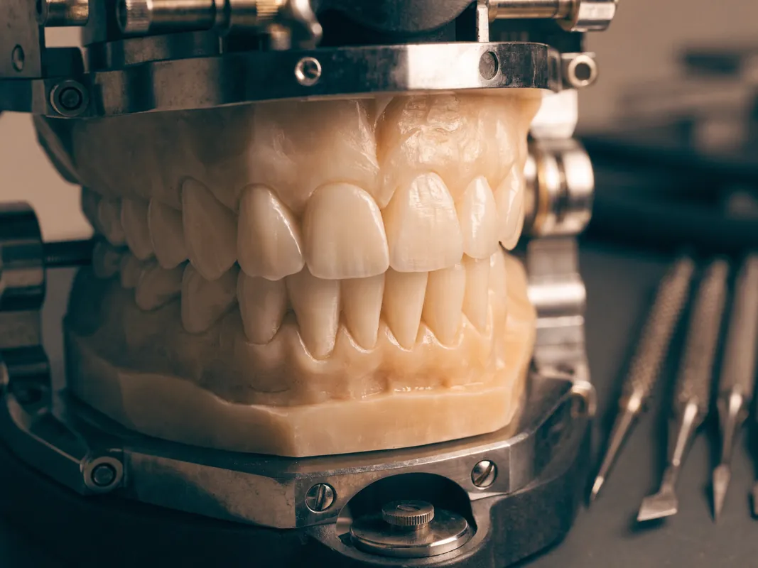
If a laboratory will produce a wax-up, model, guide, provisional or definitive restoration, the prescription should identify patient and case, tooth numbers, source records, design version, material request, shade and substrate information, contours, contacts, occlusion, surface character and any limits. The laboratory should know which decisions remain provisional and who can authorise changes.
Ask for the laboratory's legal identity and the final statement, label, invoice or conformity record appropriate to the work supplied. Design files, material labels and manufacturing records should link to the actual case. A laboratory logo in a presentation does not prove who produced the restoration.
If a technician proposes a change for manufacturability or aesthetics, the clinician must assess the clinical effect and renew patient discussion when material. Substitution should not happen silently after consent.
Consent occurs at several gates
Separate consent for collecting records, creating a simulation, trying a mock-up, performing treatment, taking marketing images and sharing data. Agreeing to one does not authorise the others.
Renew clinical consent when examination changes the proposal, before irreversible preparation, after a provisional or mock-up reveals new information, before definitive manufacture when options narrow, and before final bonding or delivery. The patient should have time to ask questions and withdraw before the relevant stage.
Consent should explain uncertainty, tissue removal, alternatives, maintenance, repair, cost, travel implications and what happens if the patient stops after an irreversible stage. A design deposit must not be described as clinical consent.
Itemise planning and treatment costs
A written quote should separate examination, photography, scanning, justified imaging, design work, models, mock-up, provisional stages, each treatment, laboratory work, materials, reviews, records and foreseeable additions. State which planning fee remains payable if treatment is declined and whether usable files are supplied.
Identify deposits, currency, taxes, expiry, cancellation, refund, revision, remake and plan-change terms. Mark estimates that remain provisional until examination. Do not hide additional teeth, gum procedures, restoration replacement or laboratory changes behind a smile package total.
Travel services should be provided under separately identifiable terms. A design should remain clinically reviewable even if transport or accommodation arrangements change.
Records and handover
Ask for copies of the clinical assessment, photographs you authorised, scans or models in an accessible format where available, design versions, shade information, consent, itemised invoice and final treatment records. Confirm whether access to proprietary files ends if treatment is declined or moved to another provider.
If treatment follows, aftercare belongs to that treatment rather than to the design itself. Obtain cleaning and maintenance instructions, review timing, warning signs and a contact route. A home dentist may need the final material and tooth-by-tooth record. Seek local care for pain, swelling, a loose temporary restoration or bite problems instead of relying on the simulation.
Health data, facial images and privacy
Photographs, scans, videos, histories and treatment proposals can reveal health and identity information. Ask which legal entity controls the data, the purpose and legal basis for each use, who receives files, whether processing occurs in another country, how access is secured, how long records are retained, and how to request access or correction.
An ordinary photograph is not automatically biometric data. Specific technical processing for unique identification can change that analysis, as the [ICO biometric guidance](https://ico.org.uk/for-organisations/uk-gdpr-guidance-and-resources/lawful-basis/biometric-data-guidance/biometric-recognition/biometric-recognition/) explains for UK-facing use. Dental and medical information can also be health data. Ask the actual controller to explain the rules that apply to the cross-border workflow.
Do not upload clinical images to a public messaging or image-generation service without a documented, appropriate pathway. A convenience tool should not receive health data merely because it produces an attractive preview.
Clinical photography is not marketing consent
Consent to take photographs for diagnosis and records is separate from permission to publish them. Ask whether images show the face, teeth only or identifying details; where they may appear; whether they will be edited; how refusal affects care; and how withdrawal works for future uses.
A treatment discount should not make marketing consent compulsory. Before-and-after images should not imply that another patient will receive the same outcome. Preserve the unedited clinical source and label simulations clearly.
If the patient permits publication, the provider should record the exact images and purposes approved. General wording such as educational use everywhere forever is difficult to evaluate and should be clarified before agreement.
File access and portability
Ask which files you can receive: original photographs, scan exports, design previews, printable models, laboratory prescriptions, material records and final treatment data. Proprietary working files may not transfer directly, but the provider should explain available standard exports, licences and any fee before payment.
Confirm whether files remain accessible if you decline treatment, change provider or request local maintenance. A screenshot is not an adequate substitute for a clinically useful scan or treatment record. Keep checksums, dates or version identifiers where the workflow supplies them.
Portability does not mean another clinician must accept the design or take responsibility for it. A receiving professional needs to assess the patient independently and decide which records are usable.
Accessibility and communication
Tell the provider about language, hearing, vision, mobility, sensory, cognitive or anxiety-related needs. Request information in a usable format, enough time for decisions, interpretation where needed, an accessible facility and permission for an appropriate support person. Confirm confidentiality when another person helps communicate.
Automated translation may support logistics but can miss clinical meaning. The named clinician should check that the patient understands options, tissue removal, uncertainty, costs and the right to stop. A coordinator should not rewrite clinical advice while translating it.
Digital design can improve explanation only when the patient can actually see, navigate and question it. Provide a non-digital route for someone who cannot or does not want to use a portal or three-dimensional viewer.
Red flags in a digital smile offer
Pause if the offer relies on:
- a final plan approved from selfies alone;
- no named examining dentist or legal facility;
- a simulation presented as diagnosis;
- one fixed number of veneers before tooth assessment;
- no separation of natural teeth and restorations;
- no tooth-preservation or no-treatment option;
- software or AI presented as clinical authority;
- no version history or source records;
- hidden tooth reduction in a front-view image;
- a mock-up described as proof of final colour or function;
- no laboratory identity or material prescription;
- treatment consent bundled with photography permission;
- files withheld if treatment is declined without prior disclosure;
- unitemised clinical and travel charges;
- no local aftercare, complaint or record route.
Resolve gaps before payment or irreversible treatment. Several unresolved gaps justify an independent opinion or stopping the process.
Digital smile planning worksheet
| Decision | Evidence to obtain |
|---|---|
| Patient objective | Plain-language concern and no-treatment option |
| Oral-health readiness | Examination and defined diagnostic records |
| Clinical ownership | Named dentist for each decision and procedure |
| Capture purpose | Reason for each photo, scan, video or image |
| Source quality | Date, calibration, completeness and limitations |
| Design version | Author, inputs, revision and intended use |
| Tissue preservation | Additions, reductions and alternatives per tooth |
| Function | Speech, bite, movement, hygiene and access checks |
| Trial stage | Reversibility, criteria, feedback and stop point |
| Laboratory | Prescription, material, identity and traceability |
| Consent | Separate, continuing gates with right to withdraw |
| Data | Controller, sharing, security, retention and access |
| Quote | Itemised planning, treatment, laboratory and exclusions |
| Handover | Original and derived files plus final clinical records |
Separate patient preference from clinical recommendation
A useful smile-planning record distinguishes what the patient asked to explore from what the dentist recommends after assessment. “Longer front teeth” may be a visual preference, while the clinician may find bite, wear, speech, gum or restorative limits. The two viewpoints can be discussed without treating the preview as an instruction.
Ask the design notes to label preferences, clinical findings, unresolved questions and the final recommendation separately. This prevents an early sketch from reappearing later as if it were an approved treatment plan. If a requested appearance cannot be delivered conservatively or safely, the dentist should explain the constraint and offer a revised option or no treatment rather than silently changing the design.
Facial and dental reference lines need context
Design software can display facial midlines, interpupillary lines, lip curves and dental axes. These measurements depend on head position, camera orientation, expression, facial asymmetry and the chosen photograph. They are references for discussion, not commands that every tooth or gum line must be made mathematically symmetrical.
Ask which image and head position produced each reference and whether the same relationship appears during natural movement. A natural facial midline and dental midline may not coincide exactly, and correcting a harmless difference can require unnecessary tissue removal or orthodontic movement. The plan should show the benefit and biological cost of any proposed correction.
Map natural teeth and existing restorations before redesign
The preview should identify which visible units are natural teeth, fillings, bonding, veneers, crowns, bridge units, implant restorations or missing spaces. Their colour, position and ability to be altered differ. A screen can recolour all units together even though whitening affects natural teeth differently from restorations.
Ask for a tooth-by-tooth map with condition, material and planned action. “Keep,” “repair,” “replace,” “move,” “whiten,” “add,” “reduce” and “monitor” should not be blended into one cosmetic layer. Existing work may be clinically sound even if it does not perfectly match a new target. Replacing it requires its own diagnosis, tissue implications, quote and consent.
Multi-treatment sequences need explicit dependencies
A smile plan may combine hygiene or periodontal care, whitening, orthodontics, bonding, veneers, crowns, implants or other procedures. Each step can change anatomy, shade, gum level, bite or available records for the next. A final design made before those changes may become outdated.
Ask for a dependency map: what must be diagnosed or stabilised first, which record is repeated after change, when material and shade are selected, and which stage remains optional. The digital smile design and veneers guide separates a visual proposal from irreversible restorative decisions. The patient should be able to stop after a reversible stage and receive an updated plan rather than being carried automatically through a package.
Define what the preview is intended to test
A preview can become more useful when it answers named questions: approximate tooth length, edge relationship, gap closure, shade direction, visible gum transition, lip support or another concern. It becomes less useful when it is called simply the final smile. Each version should state its purpose and limitations.
Ask how the question will be verified—through photographs, a three-dimensional model, a mock-up, a provisional restoration, speech assessment or clinical measurement. Acceptance of a front-view image should not be interpreted as approval of preparation, material, gum surgery, bite or the number of treated teeth. Those require their own records and consent gates.
Record what happens when the design cannot be delivered
Examination, preparation, tissue response, laboratory constraints or patient feedback may make the approved preview unsuitable. The plan should explain who pauses work, who proposes alternatives, which records are repeated and when the patient can reconsider. A departure date or prepaid package should not force an unsafe approximation.
If a change adds tooth reduction, treatment, laboratory work, cost or travel, require an updated written proposal and active consent. The old image should remain in the version history so the difference is visible. A revised design can be appropriate, but silent substitution undermines both communication and continuity.
Use an approval log instead of a single final screenshot
Keep dated versions showing source files, designer, clinical reviewer, patient feedback, requested changes and the intended use of each output. Mark whether a version is exploratory, selected for mock-up, accepted for provisional manufacture or superseded. This prevents a coordinator, clinician, technician and patient from working from different images.
Approval should be granular. A patient might accept general tooth length but not shade, tissue removal or a crown proposal. The record should not convert a thumbs-up message into consent for all treatments visible on screen. Final consent remains a clinician-led process with an opportunity to ask questions.
Design maintenance and repair into the proposal
The preview often concentrates on the front view, while future care depends on margins, contacts, material thickness, cleaning access, component retrievability and local repair. Ask how each proposed restoration is cleaned, examined, adjusted, repaired or replaced and what healthy tissue may be affected later.
For implant restorations, request system and component information. For tooth-supported work, request preparation, foundation, material and bonding or cementation records. If an attractive shape cannot be maintained by the patient's hands or serviced near home, compare another contour, material or treatment. Maintenance is a design input, not a footnote after delivery.
Arrange receiving-clinician review before travel
A dentist near home may be asked to monitor gums, bite, sensitivity, implant components or restorative work. Ask in advance whether they are willing to review the proposed treatments and which records they require. They must make their own clinical judgement and do not automatically adopt the digital plan.
The handover should include baseline findings, original and derived files, version history, tooth-by-tooth treatment, material and laboratory records, bite information, aftercare and the overseas clinician's contact. A screenshot cannot replace clinically useful files and notes. Continuity should be planned before irreversible work, not after a problem develops.
Quote changes need the same version control as designs
Link each written quote to a dated clinical plan and design version. When tooth count, procedure, material, laboratory, mock-up, provisional stage or travel changes, issue a revised itemisation. This helps the patient compare the financial effect of a clinical change rather than discovering it at reception. Keep the superseded quote as part of the decision record so neither side has to reconstruct what was previously authorised from memory.
Separate non-clinical planning fees from examination and treatment. State which files are supplied if the patient stops, which design revisions are included and how remakes differ from patient-requested changes. A deposit should not expire before the patient has enough information to understand an irreversible proposal.
Sources and evidence limits
This guide uses the [GDC consent standard](https://standards.gdc-uk.org/pages/principle3/principle3), [GDC record standard](https://standards.gdc-uk.org/pages/principle4/principle4), [ICO health-data guidance](https://ico.org.uk/for-organisations/uk-gdpr-guidance-and-resources/lawful-basis/special-category-data/what-is-special-category-data/), [ICO biometric guidance](https://ico.org.uk/for-organisations/uk-gdpr-guidance-and-resources/lawful-basis/biometric-data-guidance/biometric-recognition/biometric-recognition/), [ADA patient-record overview](https://www.ada.org/resources/practice/practice-management/documentation-patient-records) and [NHS dental-treatment overview](https://www.nhs.uk/live-well/healthy-teeth-and-gums/dental-treatments/).
These sources describe UK or US-facing professional, record and data questions. They do not certify a provider, software or laboratory in Turkey and do not decide which treatment is appropriate. Technology, products, laws and file formats change; verify the current provider, jurisdiction, instructions and data terms before sharing records or paying.
Travel boundary
Planning can often begin near home and may be bundled with a later clinical visit, but no travel should be booked solely on a remote preview. Compare the full written scope, allow time for in-person reassessment and preserve room in the schedule if the plan changes. The safest commercial promise is a transparent planning process; the image itself cannot promise the clinical outcome.
If veneers are one of the options under consideration, continue with the digital smile design and veneers decision guide. It separates the image, three-dimensional model, mock-up, provisional stage and definitive restoration, then maps tooth preservation, laboratory traceability, consent, quotation and aftercare.