A failed implant is a description, not a diagnosis. A patient may use the word failure for pain, a loose crown, a fractured screw, bleeding tissues, progressive bone loss, an implant that never became stable, an implant that was removed, or a result that cannot be cleaned or used comfortably. Those problems do not share one cause and they do not lead automatically to the same treatment.
This guide is general information, not personalised dental or medical advice. It does not promise that an implant can be retained, removed atraumatically, grafted or replaced. It does not set a universal waiting period, medicine regimen or number of visits. A meaningful decision requires an in-person assessment by an appropriately qualified clinician who has reviewed the history, records, tissues, prosthesis and relevant imaging.
WeCare is not the treating dental provider. Its role is limited to enquiry and referral coordination and, if separately agreed, non-clinical travel logistics only to the extent confirmed in writing. The named clinic and named clinician must own examination, diagnosis, consent, treatment, prescriptions, device selection, records, complaints and clinical aftercare. A coordinator cannot determine why an implant failed or clear a site for another operation.
The practical objective is not to defend one clinic or blame another. It is to identify what is known, what remains uncertain, what needs urgent attention, which parts may be repairable and which alternatives deserve consideration. Re-treatment can be more complex than first treatment because evidence may be missing, anatomy may have changed and the patient may already have experienced cost, pain or loss of trust. A careful second opinion should make uncertainty visible rather than cover it with a sales timeline.
Start by defining what failed
Begin with the patient's own account. Ask what changed, when it changed, whether the change was sudden or gradual, and what the patient means by failed. Record pain, swelling, bleeding, bad taste, discharge, mobility, altered bite, numbness, a chipped restoration, a loose prosthesis, food trapping, difficulty cleaning, speech concerns and changes in appearance. Record what happened before the symptoms began, including maintenance, trauma, a new restoration or another dental procedure.
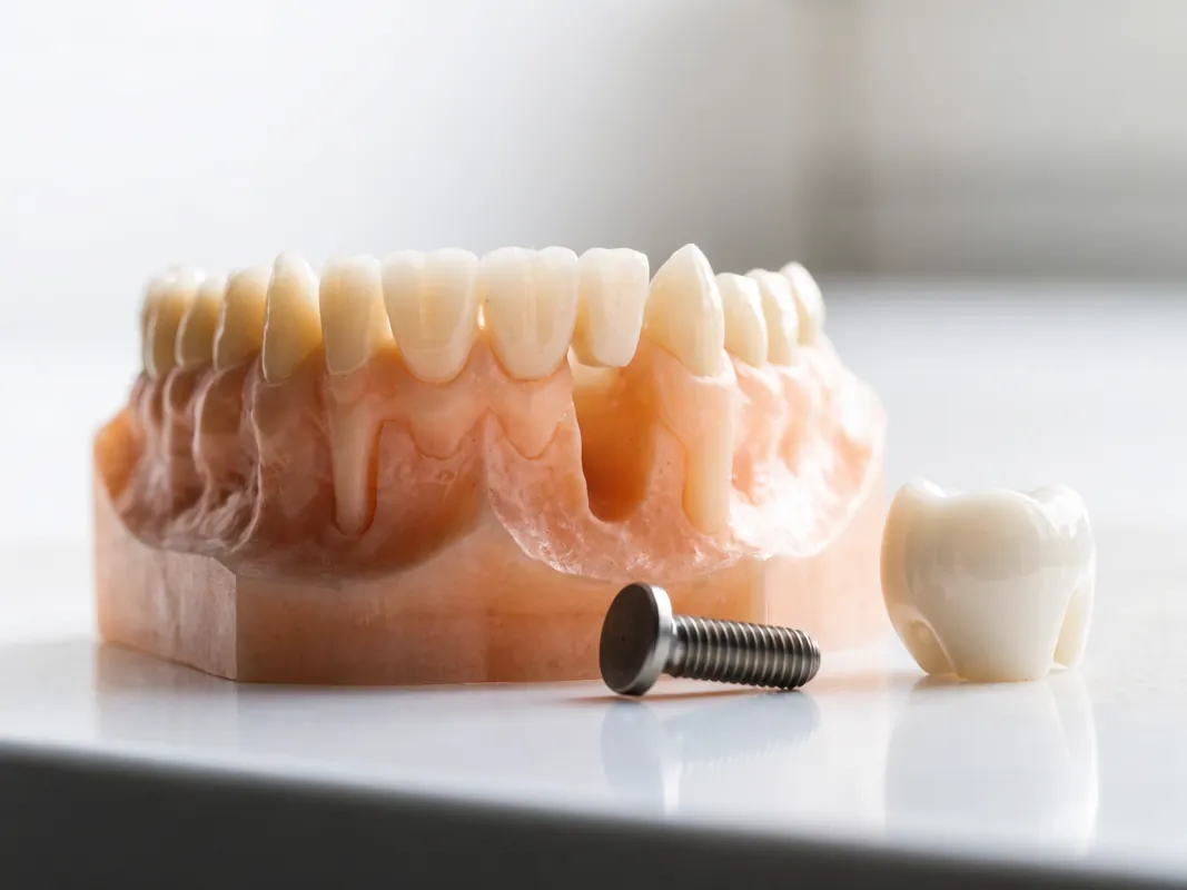
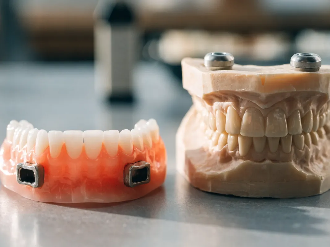
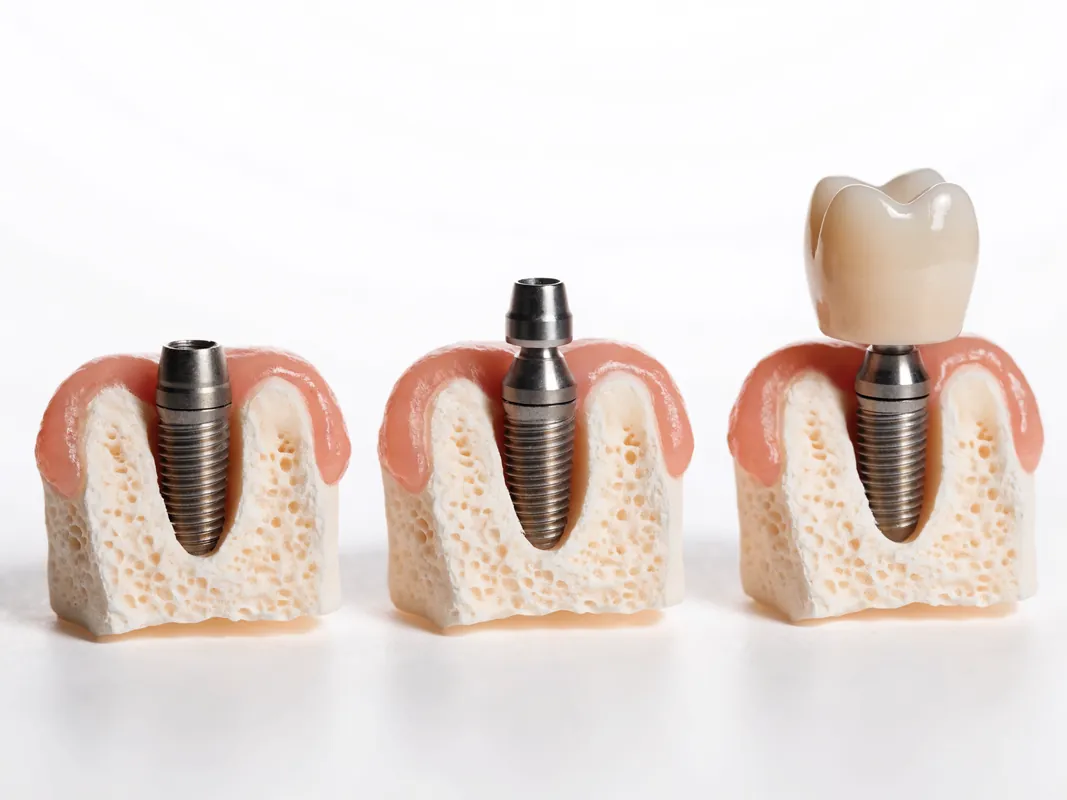
Then identify the physical level of the problem. An implant restoration is not one object. It may include the implant fixture in bone, an abutment, one or more screws, a crown or bridge, veneering material and, in a larger reconstruction, several connected components. A loose crown does not prove that the fixture has lost integration. A fractured veneering layer is different from a fractured framework. Suppuration and radiographic bone change raise a different question from a screw that has loosened without inflamed tissue.
Define whether the problem is primarily biological, mechanical, prosthetic, anatomical, functional, aesthetic or still unexplained. More than one category may apply. A prosthesis that prevents effective cleaning can contribute to inflammation. Progressive tissue loss can alter loading. A fractured component can hide a fit problem. A patient with a history of periodontitis may also have an inaccessible restoration. Labelling one factor as the cause before examination can misdirect the entire plan.
Clarify whether the implant remains in the mouth, is mobile, has already been removed, or is present but not connected to a restoration. If it was removed, ask who removed it, why, what the site looked like and whether tissue or components were retained. If the restoration is still present, avoid repeatedly testing or manipulating it at home. A clinician should decide what can be safely examined or disassembled.
The word salvage can also be misleading. It may mean retaining a treatable implant, controlling peri-implant disease, repairing or redesigning a prosthesis, removing an implant while preserving tissue, or rehabilitating the site without another implant. The patient's goal may be relief of symptoms, secure chewing, an easier-to-clean design or simply an independent explanation. Those goals should be written before procedures are proposed.
Failure stage and failure type
Timing helps organise the investigation but does not diagnose the cause. A problem identified before loading differs from a complication after the restoration has functioned. A symptom that began after a prosthetic change may warrant close examination of the restoration. An implant removed elsewhere may leave only retrospective evidence. The clinician should construct a chronology without turning timing into a universal rule.
An early stability problem may involve the implant, the bone response, local infection, surgical conditions, loading or a combination. The current clinician cannot reconstruct the original event from one radiograph or from a marketing label. Operative notes, insertion information, postoperative images, symptoms and later examinations may narrow the possibilities, but the conclusion should reflect the quality of the evidence.
A later biological problem may be peri-implant mucositis or peri-implantitis, but those terms require clinical findings. The 2018 World Workshop describes peri-implantitis as a plaque-associated pathological condition with inflammation in the peri-implant mucosa and progressive loss of supporting bone. Diagnosis uses clinical and radiographic information and is more reliable when baseline records exist. Bleeding, suppuration, probing changes and bone-level comparisons must be interpreted by a clinician; a website photograph cannot establish the diagnosis.
A mechanical event may affect the implant, abutment, screw and prosthesis at different levels. The clinician should determine which component moved or fractured, whether connected components remain retrievable, whether the interface is damaged and whether the restoration fits. Calling every loose restoration an implant failure can lead to unnecessary surgery. Conversely, repeatedly tightening a component without investigating fit, design and loading can leave the underlying problem unresolved.
An anatomical or sensory concern needs a separate pathway. Altered sensation, sinus symptoms, tissue recession, exposed threads or an implant position that compromises cleaning or restoration cannot be reduced to integration alone. The relevant clinician may need input from periodontology, prosthodontics, oral surgery, maxillofacial surgery, endodontics or another discipline. Referral is appropriate when the case is outside the examiner's competence.
An aesthetic or functional disappointment is real even when an implant remains integrated. The consent discussion should distinguish biological survival from a restoration that meets agreed functional and aesthetic goals. Re-treatment of appearance may carry tissue, component and financial consequences. The patient should not be told that a technically present implant makes every concern acceptable.
Preserve records, images and components
Evidence disappears quickly when records are not requested and removed parts are discarded. Before a new clinic dismantles or removes anything, ask what should be photographed, measured, labelled and retained. This does not mean delaying urgent care. It means preserving information whenever doing so is clinically safe.
Request the original assessment, medical history, periodontal charting, radiographs and scans, consent records, written treatment plan, surgical notes, implant placement record, component and device identifiers, laboratory prescription, statement of conformity where applicable, restoration design, postoperative instructions and maintenance records. Ask for images in their original diagnostic format when possible, not only screenshots embedded in messages.
The sequence matters. Baseline radiographs and probing measurements allow a later clinician to compare tissue and bone-level changes rather than guess from a single current image. The 2018 World Workshop specifically recommends obtaining baseline radiographic and probing measurements after completion of the implant-supported prosthesis. If those baselines are missing, the examiner should state the diagnostic limitation rather than invent a historic bone level.
Preserve invoices and written commercial terms separately from clinical records. They may identify what was sold, which components were invoiced, the promised follow-up arrangements and any stated complaint process. An invoice does not prove what was placed, but it can be compared with device documentation and clinical findings.
If a screw, abutment, crown, bridge segment or implant is removed, ask whether the treating clinician can label it, photograph it and return it or document why it cannot be returned. Infection-control, laboratory or legal considerations may affect handling. The patient should not manipulate a removed component or place it back in the mouth. The aim is traceability, not self-diagnosis.
Create a simple chronology: assessment, surgery, restoration delivery, maintenance visits, first symptom, first objective sign, interventions, medicines, component changes and current status. Mark each item as documented, patient-reported or uncertain. This prevents a later summary from presenting memory as a confirmed clinical fact.
Share records through a secure, attributable route with patient consent. Avoid posting identifiable scans and medical details publicly. The receiving clinician should confirm whether the files are complete and diagnostically usable. A coordinator may help request records, but the clinical team must decide what evidence is necessary.
Diagnosis before re-treatment
Re-treatment planning starts with a fresh diagnosis, not with copying the original plan. The new clinician should take a current medical and dental history, understand the patient's concerns, examine the whole mouth and assess the failed site in context. A single-site scan does not show periodontal health elsewhere, opposing contacts, prosthesis cleansability or all alternative treatment opportunities.
Clinical assessment may include soft-tissue appearance, plaque and deposits, bleeding and suppuration, probing where appropriate, recession, keratinised tissue, mobility, tenderness, implant and restoration position, contact points, occlusion, wear, parafunctional signs, adjacent teeth, endodontic status and the patient's ability to clean the design. Which observations are relevant depends on the presentation and the examiner's competence.
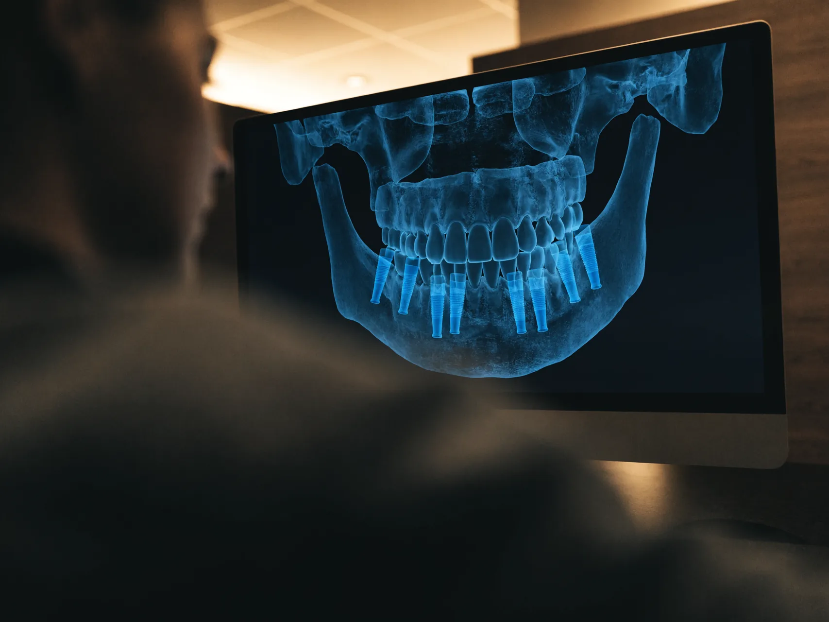
Radiographs should answer a defined question. Comparison with earlier images may show whether supporting bone has changed and how. A cone-beam scan can provide three-dimensional information when justified, but it is not a complete failure diagnosis. Image quality, artefact and radiation exposure matter. Imaging does not replace clinical examination, component assessment, periodontal charting or medical history.
If the prosthesis blocks examination or hygiene access, an appropriately skilled clinician may consider whether it can be removed or modified safely. The EFP S3 guideline notes the role of prosthesis access in peri-implant disease management. Dismantling itself can damage components or reveal that replacement parts are unavailable, so the implications should be discussed before work begins.
The differential diagnosis should remain open. Pain near an implant may arise from adjacent teeth, soft tissue, a mechanical complication, sinus disease, neuropathic pain, trauma or another condition. Bone change near an implant can have more than one explanation. A persistent or unusual lesion needs appropriate assessment and referral rather than automatic attribution to peri-implantitis.
Microbiological, laboratory or medical tests should not be sold as a standard package. A clinician should order a test only when the result can answer a relevant question and alter management. The patient should know who interprets it. A generic panel cannot determine whether a site is ready for another implant.
At the end of assessment, the written output should separate confirmed diagnoses, suspected contributors, exclusions, missing evidence and next steps. It should say whether the implant appears treatable, maintainable under observation, mechanically repairable, in need of specialist evaluation, or likely to require removal. It should also state when no responsible conclusion can yet be reached.
Biological and periodontal factors
Peri-implant tissue health is part of whole-mouth periodontal health. Examine natural teeth, periodontal stability, plaque control, smoking exposure, maintenance history and the design's cleansability. Treating one implant while active periodontal disease or inaccessible plaque-retentive contours remain unaddressed may leave important risk factors in place.
Peri-implant mucositis and peri-implantitis are not interchangeable. Mucositis involves inflammation in the peri-implant mucosa without the progressive supporting-bone loss that defines peri-implantitis. The World Workshop notes that bleeding on gentle probing is a main clinical characteristic of mucositis. The clinician should apply accepted case definitions and compare with available baseline data.
The 2023 EFP S3 clinical practice guideline describes a staged pathway for diagnosed peri-implantitis. It begins by deciding whether the affected implant is treatable. When it is, care proceeds sequentially, with an initial non-surgical step, re-evaluation and, where indicated, a surgical step, followed by supportive peri-implant care. The guideline also stresses that outcomes vary with patient, local, defect, implant and prosthetic factors. That is the opposite of a universal salvage promise.
Inflammation control includes more than a procedure performed to the implant surface. The patient must be able to clean the restoration, and professional monitoring must be feasible. Prosthesis contours, access to hygiene aids, residual cement or other local factors may matter. The clinician should demonstrate an individual cleaning method and verify that the patient can use it rather than hand over generic instructions.
Suppuration, increasing probing measurements, recession and radiographic change require clinical interpretation. Absence of pain does not prove health. Presence of bleeding alone does not prove that an implant must be removed. Trend data and the total examination matter.
If periodontitis was the reason natural teeth were lost, ask how it was treated and maintained. A history of periodontitis is relevant to risk discussion and long-term supportive care. It should not be used to blame the patient or to make a blanket decision without current findings.
When an implant has already been removed, inspect the healing site, neighbouring structures and remaining disease. Do not assume that removal eliminated the original biological problem. The treatment plan may need disease control and review before any reconstruction decision. The appropriate sequence is individual and should not be reduced to a website calendar.
Biomechanical and prosthetic factors
Mechanical and prosthetic complications require component-level investigation. Determine whether mobility comes from the fixture, an abutment connection, a screw, a cemented unit, a framework or another part. Identify wear, fracture, misfit, loss of retention, damaged interfaces, chipping and areas that prevent cleaning. Document findings before altering the assembly.
The implant system and exact component identifiers matter because connections and instruments are not universally interchangeable. A clinician should not improvise compatibility from appearance. If identifiers are unavailable, retrieval and repair may be more uncertain. Radiographs, operative records, component markings, laboratory information and the original provider may help, but the conclusion should be documented as confirmed or provisional.
Occlusion and loading should be assessed without claiming that one contact caused the failure. Look at the prosthesis design, span, cantilever, contact pattern, opposing dentition, wear, fracture history and patient-reported function. Biomechanical explanation often requires several observations. Bruxism can be relevant, but a night appliance is not an automatic cure and should be recommended only after individual assessment.
A poorly cleansable design can connect prosthetic and biological problems. The EFP guideline recommends planning restorations to permit self-performed plaque control and professional monitoring. When access is inadequate, adjustment, redesign or removal of the prosthesis may be considered by the treating clinician. The consequences for structure, appearance, cost and future repairability belong in consent.
Retaining an implant does not always mean retaining the same prosthesis. A repair, new screw, new abutment, redesigned crown or bridge, shortened span, removable solution or different overall rehabilitation may be considered. Each option has dependencies: component availability, implant position, tissue health, available restorative space, patient dexterity, maintenance access and cost.
Conversely, replacing a restoration does not treat active peri-implant disease by itself. Clinical diagnosis and disease control remain separate. A visually improved crown can still be difficult to clean; a stronger material can still be poorly designed. Material labels should not substitute for a treatment rationale.
Ask the clinician to show, on a model, image or diagram, which part is believed to have failed and which part is proposed for treatment. The written plan should name whether the fixture is retained, whether the prosthesis is reused, which components need retrieval and what happens if a component cannot be removed. This turns a vague redo into a sequence the patient can understand.
Systemic and behavioural context
The current medical history matters even when the original problem appears mechanical. Record diagnoses, allergies, medicines, previous surgery, bleeding history, immune status, diabetes care, bone-active medicines, smoking or nicotine exposure and any change since the first implant. The dentist should coordinate with the appropriate prescriber when a medical question affects planned care. A patient should not stop or alter medicine because of online content.
Behavioural factors should be discussed without moral judgement. Oral hygiene technique, access limitations, maintenance attendance, tobacco or nicotine use, alcohol, diet, clenching and the ability to attend follow-up may affect options and aftercare. The goal is to design a plan the patient can actually maintain, not to assign fault retrospectively.
If the restoration was impossible to clean with the recommended aids, document that design problem. If the patient was not given maintenance instructions, document the gap. If maintenance was offered but inaccessible because of geography or cost, that belongs in the future plan. Prevention depends on a workable system shared by patient and professionals.
Systemic factors rarely explain a local event on their own. A diagnosis such as diabetes does not prove why one implant failed. Equally, ignoring current medical status because the first surgery happened elsewhere is unsafe. The named clinician should distinguish general risk context from a demonstrated local cause.
Mental wellbeing and trust may also shape consent. A patient who has experienced pain, unexpected costs or conflicting explanations may need more time and an independent opinion. Pressure created by a deposit, flight or expiring offer can undermine a careful decision. The patient should be free to pause, seek records and decline another implant.
Urgent triage and emergency boundary
Elective re-treatment should not delay urgent care. Worsening swelling, discharge, severe pain, uncontrolled bleeding, fever or feeling systemically unwell needs prompt local assessment. A loose restoration that risks being swallowed or inhaled also needs timely professional advice. Do not repeatedly tighten, cut or remove components at home.
NHS urgent dental guidance advises seeking urgent dental care for severe or persistent mouth pain, enlarging swelling or lumps, ongoing post-extraction pain or bleeding, and broken or loose restorations. In England, NHS 111 can direct a person who cannot access a dentist. Local pathways differ outside the UK, so identify the appropriate service where the patient is physically located.
Call emergency services for heavy mouth bleeding that will not stop, serious facial or jaw injury, or severe swelling affecting breathing, the throat, neck or eyes. A remote coordinator is not an emergency service. A photograph or video cannot assess airway risk, vital signs, spreading infection or all causes of severe pain.
Urgent treatment may be stabilisation, drainage, management of a loose part, pain control, treatment of another tooth or referral. It is not necessarily implant removal or replacement. Antibiotic prescribing, when clinically indicated, belongs to an authorised prescriber who has assessed the patient; there is no universal antibiotic rule for every suspected implant problem.
Ask the urgent clinician to document findings, treatment, medicines, radiographs and what follow-up is required. That record can later inform the definitive assessment. If the urgent clinician and planned overseas clinician disagree, elective travel should pause until clinical responsibility is clear.
Observation, removal and alternative routes
The first decision is not which new implant to buy. It is whether the current implant or restoration is treatable, maintainable, repairable or harmful to retain. The EFP pathway explicitly begins with a treatability decision for peri-implantitis. Structural, positional and prosthetic problems need equivalent case-specific judgement.
Observation may be reasonable only when the named clinician has defined what is being observed, why intervention is not currently preferred, which baseline measurements will be used and what change would trigger action. Observation is active monitoring, not abandonment. It requires a maintenance route the patient can access.
Retaining the fixture may be considered when the diagnosis and tissue status support it and the prosthetic problem can be addressed. Options could involve repair, component replacement, prosthesis redesign, hygiene access or peri-implant disease treatment. The patient should understand the uncertainty, future maintenance and what would make retention unsuccessful.
Removal may be considered when an implant is mobile, structurally damaged, positioned incompatibly with a responsible restoration, associated with disease that is not treatable or maintainable, or when another clinical reason supports removal. This list is not a patient instruction. The method, risks and likely tissue consequences depend on integration, implant design, anatomy, defect and clinician competence.
Removal does not create a promise of later reimplantation. The site may be rehabilitated with a tooth-supported option, removable prosthesis, modified existing restoration, another implant configuration after assessment, or no replacement. Preserving a functional space or choosing no further surgery can be reasonable depending on goals and oral findings.
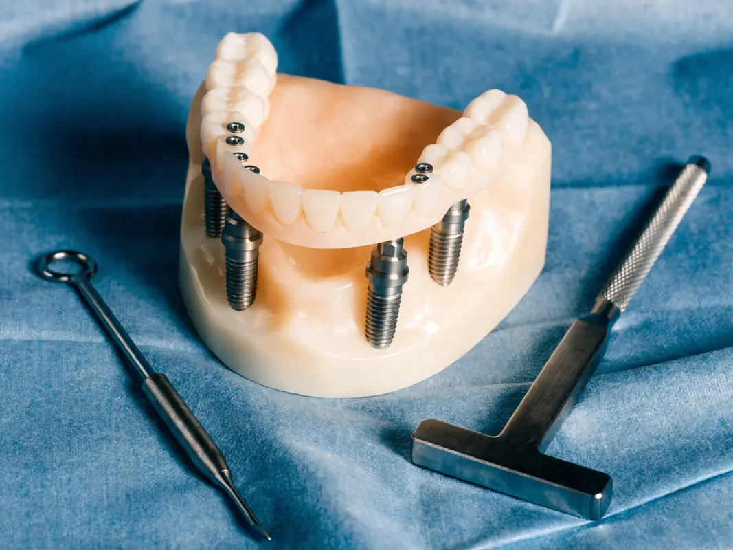
For a larger bridge or full-arch reconstruction, one problematic implant does not automatically dictate the same action for every implant. The clinician should map each fixture, component, tissue site and prosthetic dependency. A change to one support can alter the whole design, so contingency plans and costs need to be explicit.
The no-treatment alternative belongs in consent. It should include the likely practical consequences of monitoring, symptoms, function and future care, without exaggerating risk to push surgery. The patient can choose a less invasive route even when an implant option exists.

Grafting and staging uncertainty
Bone loss seen after an implant problem does not answer whether grafting is necessary, feasible or desirable. The clinician must first define the defect, tissue condition, infection or inflammation status, anatomical limits, prosthetic goal and alternatives. Imaging supports that assessment but does not make the decision alone.
A defect may reflect disease progression, removal-related change, original anatomy, implant position or more than one factor. Record what is observed and what is inferred. A statement that the site needs bone should specify where, for what proposed reconstruction and on what clinical evidence.
Simultaneous removal and reconstruction, staged site management, healing without grafting, or abandoning reimplantation may each be considered in different circumstances. The choice depends on findings at examination and sometimes findings during surgery. Consent should therefore include a primary plan, reasonable alternatives and an intra-operative contingency rather than a guaranteed sequence.
The source and type of graft material, membrane or fixation should be discussed only if clinically proposed. Ask why it is recommended, what alternatives exist, what additional surgery it involves, what documentation will be supplied and how complications would be managed. Do not infer material quality from price or a brand label.
Healing cannot be scheduled from a generic chart. Inflammation control, tissue response, surgical extent, medical context and the restorative plan all matter. The clinician should define what clinical and radiographic findings are needed before a later stage. A travel booking should follow that review, not substitute for it.
The possibility that the site remains unsuitable for another implant after removal or reconstruction must be stated before treatment. So must the possibility that the patient decides not to continue. Costs incurred at each stage and the commercial consequences of stopping should be written separately from clinical consent.
Second opinion and named clinical ownership
A second opinion should be genuinely clinical. Verify the regulator or professional register where applicable, the clinic's legal identity, the clinician's name and scope of practice, and who will perform each proposed stage. Ask whether specialist referral is needed for periodontal disease, component retrieval, nerve symptoms, extensive defects or complex prosthetic reconstruction.
Send enough evidence for useful triage, but treat remote review as provisional. Images and records can help identify missing information and urgency. They cannot reproduce palpation, mobility testing, periodontal assessment, occlusal examination or every diagnostic image. A remote quotation should state which elements may change after examination.
The clinician giving the second opinion should explain whether they agree with the original diagnosis, disagree, or cannot conclude because evidence is missing. They should distinguish facts from hypotheses and identify which additional step could reduce uncertainty. Certainty performed for a sales conversation is not a substitute for evidence.
Clinical ownership must remain with the named clinic and named clinician. The person diagnosing should be identifiable. The clinician performing surgery should confirm the plan and consent, even if another professional reviewed records remotely. The restorative clinician should understand the surgical and component plan, and the aftercare provider should receive a usable handover.
If several clinicians are involved, map responsibilities: who removes the prosthesis, who treats peri-implant disease, who removes the implant if needed, who provides grafting, who restores, who handles urgent care and who monitors long term. The patient should not be left to broker a clinical disagreement through a salesperson.
An independent local dentist may offer a useful comparison, particularly when travel is proposed. Local care may simplify examination of symptoms, retrieval of old records and long-term maintenance. Choosing the local opinion or declining re-treatment is a valid outcome.
Consent without an automatic reimplantation promise
There is no automatic reimplantation. Consent must follow diagnosis and remain valid as information changes. GDC Principle 3 requires relevant options, risks, benefits and costs to be explained and treats consent as an ongoing process rather than a signature collected once.
The discussion should cover retaining or treating the existing implant, repair, removal, reconstruction, another prosthetic route, postponement and no treatment. For each option, explain what is known about the cause, what remains uncertain, which extra procedures may become necessary, how appearance and function may change, and what maintenance is required.
Re-treatment risk is site-specific. It may include pain, swelling, bleeding, infection, tissue recession, additional bone or soft-tissue loss, damage to adjacent structures, sensory change, sinus involvement, component fracture, prosthetic compromise, need for further procedures and the possibility that the desired restoration cannot be delivered. The named clinician should tailor the discussion to the actual anatomy and plan.
Consent must include uncertainty about timing and travel. A proposed sequence can change after clinical examination, removal of a prosthesis, review of records or surgical findings. The patient should know who makes that decision, what evidence supports it and what happens financially if the plan changes.
Language support should be independent enough for the patient to ask questions and should preserve confidentiality. Do not assume a companion can interpret complex consent. The patient should receive the written plan in a form they can understand and have time to compare it with another opinion.
Consent can be withdrawn before a procedure. A deposit or travel booking does not make clinical consent irrevocable. If the patient feels pressured, does not understand the plan, or has not met the operating clinician, pause.
Records, materials and component traceability
GDC Principle 4 describes complete, accurate and contemporaneous records and identifies radiographs, photographs, models, consent forms, laboratory prescriptions, statements of conformity and referral letters as parts of the patient record where available. A failed-implant investigation particularly depends on that continuity.
Before treatment, document the diagnosis, findings, comparison images, medical history, options, decisions, responsible clinicians and estimated costs. During treatment, record the procedure, site, anaesthesia, prescribed medicines, components removed or placed, unexpected findings and any departure from the plan. After treatment, provide the patient with a clear handover.
Component traceability should identify the implant system and relevant device or component references when known. The record should distinguish confirmed identifiers from guesses. If an unknown connection limits repair, say so before dismantling. A compatible-looking part should not be represented as verified compatibility without appropriate evidence.
The patient should receive copies of relevant device documentation, procedure records, diagnostic images, laboratory information, maintenance instructions and referral correspondence. Keep the information in a format another clinician can use. A screenshot of a scan inside a chat may be helpful for discussion but is not an adequate substitute for the original diagnostic file.
If tissue is sampled or a component is sent to a laboratory, explain why, who is responsible for the result and how it will affect the plan. Do not imply that every failure needs laboratory testing. The question should drive the test.
Data transfer between countries adds privacy and continuity questions. Ask where records are stored, who receives them, how consent is recorded and how corrections or access requests are handled. Share only what is necessary through an appropriate route.

Local aftercare and handover
Arrange local aftercare before elective re-treatment, not after a complication. Ask whether a local dentist is willing and equipped to review the planned work. Another clinician is not automatically responsible for treatment performed elsewhere and may not have compatible instruments or components.
The handover should include diagnosis, procedures, sites, components, images, medicines, expected findings, individual hygiene instructions, maintenance plan, urgent signs, direct clinical contact and the threshold for in-person review. It should also explain what remote advice cannot assess.
Supportive peri-implant care is not optional background. The EFP guideline describes it as essential for preserving peri-implant tissue health. The interval and methods should be individual, based on tissue condition, prosthesis design, risk factors and the patient's ability to attend. This guide does not set a universal schedule.
At follow-up, compare clinical findings with a meaningful baseline. Monitor tissue inflammation, probing changes where appropriate, suppuration, recession, bone levels when indicated, prosthesis integrity, occlusion, hygiene access and patient-reported function. A stable implant still needs maintenance; a treated peri-implantitis site remains a site with a disease history.
Remote images can support communication but cannot replace examination when symptoms persist or worsen. The patient should know which clinician to contact locally, which service handles urgent care and what information to bring. If the treating clinic offers remote review, its hours, limitations and escalation route should be written without presenting it as emergency coverage.
If the patient cannot access reliable long-term maintenance, that may change whether a complex implant route is sensible. A simpler removable or tooth-supported plan may provide a more maintainable option. Clinical planning should account for real life, not only what can be performed during one trip.
Travel and the no-travel option
Travel adds discontinuity, time pressure and cost to an already uncertain re-treatment case. It can separate the diagnostic clinician from the urgent-care provider and the surgeon from the long-term maintenance clinician. Those risks do not make all overseas care inappropriate, but they must be considered explicitly.
Do not book travel from a remote promise that a site can be reimplanted. Ask what must be assessed in person, what may change, who has reviewed the original records and whether the operating clinician has accepted the case. The written plan should separate a provisional remote view from the definitive diagnosis.
Travel logistics must be confirmed in writing and kept separate from clinical care. Clarify dates, change conditions, companion needs, accessibility, local transport, accommodation, who pays when clinical timing changes and whether a longer stay might be needed. Do not infer a travel service from a treatment headline.
Discuss the return journey with the treating clinician. The relevant question is not a universal number of days; it is which examination findings must be satisfactory, what symptoms require delay and who will provide care if a problem appears during transit or after return.
Insurance may exclude planned dental care, existing complications, extra accommodation, changed flights or treatment abroad. Ask the insurer directly and retain the response. A clinic or coordinator should not promise coverage on an insurer's behalf.
The no-travel option should remain visible. A local second opinion, local disease control, local prosthetic repair, local removal, a removable alternative or no further implant surgery may reduce continuity problems. A hybrid approach may use local assessment and maintenance even if another clinician later performs a defined procedure. Choosing not to travel is a legitimate clinical and personal decision.
Itemised quote and remedial commercial terms
Commercial terms must remain separate from the clinical diagnosis and consent. A clinician decides what is indicated; a written agreement explains price, inclusions, exclusions, cancellation, complaints and any commercial response if treatment does not proceed as planned. One must not be used to distort the other.
Ask for an itemised estimate by diagnostic, surgical, grafting, component, prosthetic, laboratory, medicine, sedation, imaging and follow-up elements that are actually proposed. Mark provisional items and state what finding would activate them. Clarify what happens if the existing prosthesis cannot be removed, a component is unavailable, the site is unsuitable for the proposed reconstruction or the patient chooses an alternative.
Read remedial commercial terms before paying. Ask who decides whether further work is clinically necessary, whether an independent assessment is accepted, what records must be supplied, whether return to the original clinic is required, which professional fees are covered, which travel costs are excluded and which time or maintenance conditions apply. Do not reduce these terms to a single reassuring label.
A commercial commitment cannot guarantee biological healing, eliminate a complication or compel a clinician to provide inappropriate treatment. It also should not remove the patient's right to complain. Verify the clinic's legal entity, regulator where applicable, written complaint process and available independent route.
GDC Principle 2 requires a written treatment plan with proposed treatment and a realistic indication of cost, with changes communicated in writing. For overseas care, check the standards and complaint route in the treating jurisdiction. The GDC's patient guidance on going abroad recommends checking qualifications, regulation, aftercare and what happens if something goes wrong.
Keep invoices, receipts, written quotations, consent records and clinical records. If a disagreement develops, describe the chronology and requested resolution factually. Do not discard components or alter records in an attempt to prove a claim.
Questions to ask at a failed-implant consultation
About the diagnosis
- What exactly has failed: the fixture, tissue, abutment, screw, crown, bridge or another part?
- Which findings are confirmed and which are hypotheses?
- Does the site meet a recognised case definition for peri-implant disease?
- What baseline records are available for comparison?
- Could an adjacent tooth, sinus, nerve or another condition explain the symptoms?
- Is the implant treatable, maintainable, repairable or indicated for removal, and why?
About records and components
- Have you reviewed the original surgical and restorative records?
- Is the implant system confirmed?
- Are compatible instruments and components available?
- What will be photographed and recorded before dismantling?
- Can removed components be labelled and retained or fully documented?
- Which original image files do you need?
About options
- Can the existing fixture be retained?
- Can the prosthesis be repaired or redesigned?
- What non-implant alternatives are reasonable?
- What does observation involve and what triggers intervention?
- Why is removal proposed?
- Is grafting an option, a requirement for this particular plan, or still uncertain?
- What could make later reimplantation inappropriate?
- What happens if I choose no further surgery?
About ownership and follow-up
- Who is the named clinician responsible for diagnosis?
- Who will perform each procedure?
- Who restores the implant and who maintains it?
- Which local clinician will review me after I return?
- What can remote review not assess?
- Where do I seek urgent care?
- How will my records be transferred securely?
About cost and travel
- Which parts of the quote are fixed and which are provisional?
- What extra cost could arise after dismantling or surgery?
- What happens commercially if the plan changes or stops?
- What do the complaint and remedial terms require?
- Which travel services, if any, are separate and confirmed in writing?
- What is the local or no-travel alternative?
Failed-implant planning checklist
Before seeking a second opinion
- Write a symptom and treatment chronology.
- Request the complete record and original diagnostic image files.
- Request implant and component identifiers.
- Gather consent forms, treatment plans, laboratory documents and invoices.
- List current health conditions, allergies and medicines.
- Photograph visible changes without manipulating the site.
- Identify urgent symptoms that need local care now.
- Define what you want from the second opinion.
Before accepting investigation or dismantling
- Verify the clinic and named clinician.
- Confirm the clinical question each test or image will answer.
- Ask what could be damaged or become irretrievable during dismantling.
- Agree how removed components will be documented.
- Obtain an itemised diagnostic estimate.
- Understand privacy and record-sharing arrangements.
- Keep remote conclusions labelled provisional.
Before accepting re-treatment
- Obtain the written diagnosis and uncertainties.
- Compare retention, treatment, repair, removal, alternative prosthesis and no-treatment routes.
- Review the role of periodontal and peri-implant disease control.
- Ask how prosthesis design supports cleaning and monitoring.
- Review medical and behavioural factors without changing medicine yourself.
- Obtain a primary plan and contingency plan.
- Confirm the clinicians responsible for surgery, restoration and aftercare.
- Read consent, itemised costs, cancellation, complaint and remedial terms.
- Arrange local maintenance and urgent access.
Before travel
- Confirm the operating clinician has reviewed the case.
- Confirm what remains undecided until examination.
- Keep travel services separate from clinical consent.
- Check insurance directly.
- Plan for changed dates and extra local care.
- Carry records and medicine information securely.
- Identify urgent dental and emergency services at the destination.
- Keep the no-travel option open until the clinical plan is clear.
Before discharge
- Receive the final diagnosis and procedure record.
- Receive component and material documentation.
- Obtain relevant images in a usable format.
- Receive individual aftercare and hygiene instructions.
- Confirm urgent signs and direct clinical contact.
- Confirm the local handover and review plan.
- Obtain the updated itemised invoice and complaint route.
- Ask which findings must be satisfactory before travel.
During long-term maintenance
- Attend the individually agreed local review.
- Keep the restoration clean using demonstrated methods.
- Report new mobility, swelling, discharge, pain, bleeding or functional change.
- Keep clinical and radiographic comparisons.
- Ensure another clinician can identify the components.
- Revisit the plan if health, medicine, dexterity or access to care changes.
Sources and review dates
Guidance and evidence develop. The named treating clinician should check the current versions and apply them to the individual patient. This guide used:
- EFP S3 clinical practice guideline, Prevention and treatment of peri-implant diseases, published 2023 and accessed 29 August 2026: diagnosis, treatability decisions, sequential disease management, prosthesis access and supportive peri-implant care. https://onlinelibrary.wiley.com/doi/full/10.1111/jcpe.13823
- World Workshop consensus report, Peri-implant diseases and conditions, published June 2018 and accessed 29 August 2026: definitions of peri-implant health, mucositis and peri-implantitis, and the importance of baseline radiographs and probing. https://pubmed.ncbi.nlm.nih.gov/29926955/
- SDCEP Prevention and Treatment of Periodontal Diseases in Primary Care, second edition, published November 2025 and accessed 29 August 2026: risk assessment, peri-implant monitoring, primary-care management and referral boundaries. https://www.periodontalcare.sdcep.org.uk/media/vjgfn5ak/sdcep-prevention-and-treatment-of-periodontal-diseases-in-primary-care-2nd-edition-nov-2025.pdf
- GDC Principle 2, Communicate effectively with patients, accessed 29 August 2026: written treatment plans, realistic cost information, changes and emergency arrangements. https://standards.gdc-uk.org/pages/principle2/principle2
- GDC Principle 3, Obtain valid consent, accessed 29 August 2026: relevant options, risks, benefits, costs and continuing consent. https://standards.gdc-uk.org/pages/principle3/principle3
- GDC Principle 4, Maintain and protect patients' information, accessed 29 August 2026: complete records, radiographs, photographs, laboratory documents and referral handover. https://standards.gdc-uk.org/pages/principle4/principle4
- GDC, Going abroad for dental treatment, accessed 29 August 2026: provider verification, full medical history, aftercare and complaint planning. https://www.gdc-uk.org/standards-guidance/information-for-patients-public/going-abroad-for-dental-treatment
- NHS urgent dental guidance, accessed 29 August 2026: urgent dental access and emergency signs including uncontrolled bleeding and severe swelling affecting breathing. https://www.nhs.uk/nhs-services/dentists/how-to-find-an-nhs-dentist-in-an-emergency/
The evidence supports diagnosis, staged decisions and maintainable aftercare. It does not support an automatic promise that every failed implant can or should be replaced.