A missing premolar or molar creates a decision, not an automatic order for an implant. The useful starting questions are whether a damaged tooth can be predictably retained, whether the space needs replacement for this patient, which alternatives preserve the most healthy tissue, and whether the chosen restoration can be cleaned, loaded and maintained over time. The answer can differ between an upper first premolar, a lower first molar and a last tooth at the end of a shortened dental arch.
Posterior implant planning is both biological and restorative. Bone and soft tissue must be assessed, but the proposed crown position should guide the surgical plan. The maxillary sinus may limit available height in the upper posterior jaw. The inferior alveolar nerve, mental foramen and lingual undercut may be relevant in the lower jaw. Space, opposing teeth, adjacent roots, tooth movement, bite, wear, bruxism, hygiene and local aftercare can change the choice even when an implant would physically fit.
This guide is educational information for adults researching a back tooth dental implant, molar implant or premolar replacement, including people considering treatment abroad. It cannot diagnose a tooth, interpret a scan for an individual, prescribe surgery or promise a result. The named treating clinician must examine the mouth, determine whether the tooth is restorable, justify any imaging, compare reasonable options, obtain valid consent and remain responsible for treatment and aftercare.
For cross-border care, identify the named legal provider, named dentist, clinic address, professional registration route, direct clinical contact, complaint process and indemnity or insurance arrangements before sharing health information or paying. Verify those details independently and keep travel logistics separate from clinical consent.
Preserve a restorable back tooth before discussing replacement
An implant replaces a missing or non-restorable tooth; it does not improve a tooth merely by replacing it. If the premolar or molar is still present, the first decision is whether it can be retained with a reasonable prognosis and maintenance burden. This may require restorative, endodontic, periodontal or combined assessment. A coordinator and a panoramic image alone cannot declare a tooth hopeless.
The American Association of Endodontists position statement says retention should be the first consideration for a compromised natural tooth and that implants should not be treated as a cure-all. This is not a rule that every tooth must be saved. Some teeth have a fracture pattern, caries extent, periodontal loss, resorption, structural deficiency, infection or strategic problem that makes extraction reasonable. The point is to document why retention is or is not realistic before an irreversible extraction.
Ask the clinician to record:
- the diagnosis for the tooth and surrounding tissues;
- whether pain comes from the tooth, gums, bite, sinus, muscles or another source;
- the remaining sound tooth structure and whether a durable restoration is feasible;
- pulp and apical findings, including whether endodontic treatment, retreatment or specialist review is relevant;
- periodontal support, mobility, furcation involvement and the ability to maintain the tooth;
- cracks or fractures and how confidently their extent can be established;
- existing crown, post, filling or root-canal quality;
- the alternatives, uncertainties, likely maintenance and costs of retention;
- the reasons extraction is recommended if retention is rejected.
Comparisons between endodontically treated teeth and implant restorations use different definitions of success, survival and complications. Recent systematic reviews do not establish a universal winner. Both pathways can require further treatment. A written patient decision should weigh diagnosis, complication burden, tissue preservation, time, cost, preference and local access rather than using one headline percentage.
If the assessment is uncertain, seek an opinion from a clinician with relevant restorative, endodontic or periodontal expertise before extraction. After the tooth is removed, the retention option is gone.
Decide whether the missing tooth needs replacement
Not every missing posterior tooth must be replaced immediately, and not every untreated space is harmless. The decision depends on which tooth is absent, how many functional contacts remain, whether there is an opposing tooth, the state of adjacent teeth, the patient's chewing experience, diet, speech, comfort, appearance, disease risk and expected future care.
The shortened dental arch concept recognises that some people can function acceptably without replacing every missing molar. Clinical research comparing removable replacement with non-replacement in selected shortened arches has not established one strategy as universally preferable. A 2018 systematic review found insufficient evidence to recommend one tooth-replacement strategy for all adults with reduced dentitions. This supports shared decision-making, not neglect.
Replacement may be more compelling when the patient reports impaired chewing, food trapping, recurrent trauma, instability of an existing prosthesis or a functional problem linked to the space. It may also form part of a larger plan to maintain a stable restoration, provide a strategic support or protect a vulnerable remaining dentition. Conversely, a last molar without an opposing contact may add limited benefit for some patients, while surgery and maintenance still carry burdens.
Ask what would happen if the space were monitored rather than restored. The clinician should discuss the observed position of neighbouring and opposing teeth, the actual occlusal scheme, caries and periodontal risks, symptoms and the feasibility of future replacement. “All gaps always cause collapse” is too absolute. Research on bounded posterior spaces found that adjacent-tooth movement was often gradual and minor after the earlier post-extraction period, although individual movement varies. Observation should therefore be active and documented, not assumed safe forever.
No immediate replacement can be a legitimate option when benefits do not justify intervention, when disease needs stabilisation first, when medical or anatomical questions remain, or when the patient chooses to defer. Record the review trigger: new chewing difficulty, change in tooth position, overeruption, food trapping, fracture, caries, periodontal change or a future restorative need.
Premolars, first molars, second molars and wisdom teeth are different sites
“Back tooth” is not one anatomical or functional category. A premolar may be visible in the smile and may contribute to canine-premolar guidance or bridge support. A first molar often provides a substantial functional contact. A second molar lies farther back, where access and cleanability may be harder. A wisdom tooth has a different strategic value and is not routinely replaced with an implant merely because it was removed.
The clinician should identify the tooth using a recognised notation and show the patient on the chart. Clarify whether the gap is bounded by teeth on both sides or is a free-end space. Clarify whether the opposing tooth is natural, restored, implant-supported, removable or absent. The available mesiodistal space, ridge width, vertical restorative space and root positions of neighbours affect whether an implant crown can be designed without overcontour or contact problems.
For multiple missing posterior teeth, a tooth-by-tooth replacement count is not automatically the correct design. Options can include separate implants, an implant-supported bridge with pontics, a tooth-supported bridge, a removable partial denture, a shortened arch or a combination within a wider rehabilitation. The restorative clinician should explain why the proposed number and positions provide cleanability and manageable load without assuming that every absent root needs one fixture.
If the remaining dentition is extensively compromised, jumping from “several missing molars” to a full-arch extraction proposal is unsafe. Each remaining tooth needs a documented prognosis and preservation assessment. Full-arch options belong to a separate diagnosis and consent process, not a shortcut for isolated posterior spaces.
Urgent disease comes before elective implant planning
Pain, swelling, pus, fever, spreading redness, difficulty opening the mouth or a bad taste may indicate infection requiring local assessment. A fractured tooth can have sharp edges or pulp involvement. New numbness, altered sensation or sinus symptoms also require diagnosis. Do not wait for a travel quotation when urgent care is needed.
An abscess is not treated by scheduling a future implant. The source must be identified and managed. Antibiotics are not an automatic substitute for drainage or dental treatment, and they should not be prescribed by a coordinator. The responsible clinician must consider allergies, interactions, antimicrobial stewardship and the actual diagnosis.
In England, NHS 111 can direct urgent dental care. Severe swelling affecting breathing or swallowing, serious facial trauma, uncontrolled bleeding or other emergency features require emergency services. Outside the UK, use the local urgent and emergency pathways. Stabilising disease can change the later replacement plan.
Build a complete posterior diagnosis
A responsible assessment connects the whole mouth to the local site. It may include:
- current medical, dental and medicine history, allergies and previous adverse reactions;
- smoking, vaping, alcohol and other relevant health behaviours;
- the history of tooth loss, extraction, infection, trauma and previous grafting;
- pain, chewing limitations, food trapping and patient priorities;
- periodontal charting and oral-hygiene assessment;
- caries, cracks, restorability and endodontic findings of adjacent and opposing teeth;
- ridge shape, keratinised or mobile mucosa, vestibular depth and cleanability;
- space between adjacent teeth, root positions and restorative height;
- opposing contacts, excursive contacts, wear, clenching, grinding and previous fracture;
- initial two-dimensional imaging where justified and review of existing images;
- cross-sectional imaging for presurgical implant planning when indicated by current guidance;
- a diagnostic restoration, scan, model or wax-up that defines the intended crown before implant selection;
- local aftercare capability and the patient's ability to maintain the site.
Medical history matters because an implant is elective surgery followed by a long-term restoration. Diabetes control, immune conditions, bleeding risk, osteoporosis medicines, cancer treatment, cardiovascular disease, smoking and other factors may change risk, coordination or timing. Do not stop a prescribed medicine because a dental website suggests it. The dental clinician should contact the relevant prescriber with permission when an individual medical question arises.
A photo or panoramic image may help an initial discussion, but it cannot determine restorability, soft-tissue quality, three-dimensional anatomy, bite or surgical suitability. Any remote plan must remain provisional until the named clinician completes the required assessment.
Space, neighbouring teeth and the opposing tooth
A crown needs adequate space in three dimensions. Too little mesiodistal room can produce poor contacts or an overcontoured crown. Excessive space may require an unusually wide crown, orthodontic redistribution, more than one restorative unit or acceptance of a non-standard contour. Adjacent roots may converge even when crown space looks generous. The proposed implant position must respect those roots and support a cleanable emergence profile.
Vertical restorative space also matters. An opposing tooth can erupt into an unopposed space, reducing room for a crown. Adjacent teeth can tip or rotate. These changes are variable and should be measured, not presumed from the age of the gap. Restoring the space may require orthodontic movement, adjustment or restoration of another tooth, but every added procedure needs its own indication and consent.
The opposing tooth influences function and material choice. An implant crown opposing a natural tooth is not the same load context as one opposing another implant restoration, a denture tooth or no tooth. The clinician should map centric and excursive contacts and note how contacts may change as other teeth move or wear.
Contacts between a posterior implant crown and adjacent natural teeth require monitoring. Natural teeth can move differently from an osseointegrated implant, and an open contact can develop, contributing to food impaction or gum symptoms. The patient should know how contacts will be assessed and who can adjust or repair the restoration locally.
Compare all reasonable replacement options
Preserve and restore the tooth
If the tooth remains restorable, options can include direct restoration, onlay, crown, periodontal treatment, endodontic treatment or retreatment, and combinations. The exact choice depends on diagnosis and remaining structure. Retention avoids extraction and implant surgery but can still have biological, technical and financial burdens. Ask for the expected maintenance and what findings would change the prognosis.
No immediate replacement or a shortened arch
Observation may be reasonable for a selected asymptomatic space with acceptable function and a monitoring plan. It avoids immediate procedure risks and cost, but it may leave food trapping, reduced chewing contacts or future movement. The decision should record patient priorities and review triggers.
A single implant-supported crown
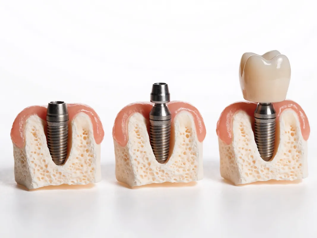
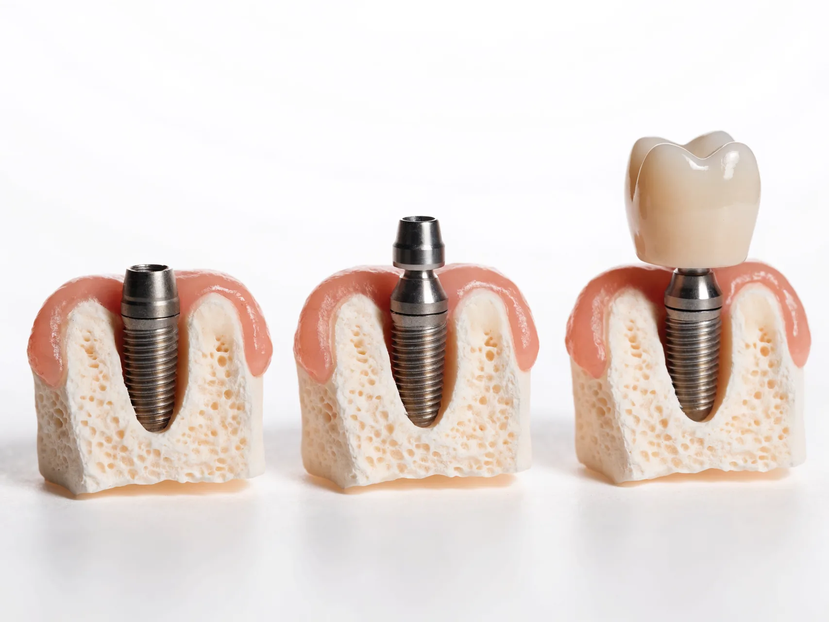
This option can replace one tooth without preparing healthy neighbouring teeth for a conventional bridge. It requires surgery, sufficient or augmentable hard and soft tissue, a restorable three-dimensional crown position, healing, a component connection and continuing peri-implant maintenance. Biological and technical complications remain possible.
A tooth-supported conventional bridge
A bridge may be relevant when adjacent teeth already need crowns or provide suitable support. It avoids implant placement at the missing site but requires preparation of abutment teeth and links their futures. Caries, endodontic complications, periodontal support, connector design and cleaning under the pontic belong in the discussion.
A resin-retained or adhesive bridge
In selected situations, an adhesive bridge can preserve more tooth tissue than a conventional bridge. Posterior load, available enamel, occlusion, span, design and operator experience affect suitability. It can debond and is not appropriate for every molar space. It should not be dismissed or promised without case-specific assessment.
A removable partial denture
A removable prosthesis may replace one or several missing teeth without implant surgery. It can restore multiple spaces and may be modified, but it introduces clasps or other components, movement, coverage, hygiene and adaptation considerations. Some patients value removability and lower surgical burden; others find it intrusive. Examine supporting teeth and tissues and explain maintenance.
An implant-supported bridge
For several adjacent missing teeth, an implant-supported bridge may use selected implant positions with one or more pontics. The design should be prosthetically driven and cleanable. The number of implants, span, connector dimensions, cantilever, material and load must be justified. It is not a simple formula based only on the number of missing teeth.
Orthodontic space closure or redistribution
In some cases, orthodontic movement may close or redistribute a posterior space, improve root position or create a safer restorative site. This requires an orthodontic assessment and may alter the whole occlusion. It is not automatically suitable or simpler, but it belongs in the alternative discussion when relevant.
Compare each option by tissue removal, surgery, expected function, cleanability, repairability, complication types, effect on adjacent teeth, time burden, local access and itemised cost. Include the no-treatment choice.
Plan from the crown backwards
An implant should not be placed wherever bone appears easiest and then handed to a restorative clinician to invent a crown. The intended occlusal surface, contacts, emergence, screw-access or cement strategy, cleansability and relationship to adjacent teeth should guide the three-dimensional implant plan. This is often described as prosthetically driven planning.
A diagnostic wax-up or digital design can help define crown volume and implant trajectory. It is a planning record, not a result guarantee. The surgeon and restorative clinician should agree the position and explain any compromise. If the ideal crown position conflicts with available bone or anatomy, alternatives can include augmentation, a different implant design, a bridge, orthodontic movement, a removable option or no implant.
Guided surgery can transfer a digital plan but does not eliminate clinical judgment or procedural error. The clinician must verify fit, mouth opening, drill access, anatomy and intra-operative findings. A guide does not make an unsuitable site suitable.
Posterior upper-jaw anatomy and the maxillary sinus
Upper premolar and molar roots may lie near the maxillary sinus. After extraction, ridge dimensions can change, and sinus anatomy varies. A sinus seen near the site does not automatically mean grafting is required. The clinician should assess the planned crown, bone volume, sinus health, residual ridge, implant options and alternatives.
Sinus symptoms, previous sinus surgery, recurrent infection or relevant imaging findings may require medical or ear-nose-throat input. Incidental mucosal thickening is not a patient-facing diagnosis by itself. The person interpreting the scan should decide whether a finding affects the dental plan and whether referral is needed.
Sinus-floor elevation can be performed through different surgical approaches, with or without simultaneous implant placement, in selected cases. The choice depends on site anatomy, residual bone, the ability to obtain stability, graft strategy, clinician competence and patient factors. This page does not set a millimetre threshold or a fixed healing period. Ask why augmentation is needed, what material is proposed, who provides it, what complications are possible and what non-graft alternatives exist.
Alternatives may include a shorter implant, different position or prosthetic design, bridge, removable replacement, orthodontic change, observation or referral, depending on the case. No technique is automatically less invasive or more successful for everyone.
Posterior lower-jaw anatomy, nerve and lingual boundaries
The inferior alveolar nerve travels within the mandible and supplies sensation to the lower lip and chin through its branches. The mental foramen and anterior loop may be relevant near premolar sites. Posterior mandibular ridges can also have a lingual undercut, where the inner surface curves inward. Two-dimensional images can obscure buccolingual anatomy.
The surgical plan must establish the relationship between the proposed implant and relevant nerve, roots and cortical boundaries. New altered sensation after surgery requires prompt direct clinical assessment. Numbness, tingling, burning or changed lip and chin sensation should not be managed only through a coordinator's messages.
Bone that looks tall on a panoramic image may still be narrow or undercut. The implant length and diameter should follow the prosthetic plan, available anatomy, system and clinician's judgment; a back-tooth label does not mandate a wide fixture. Bigger is not automatically safer or more durable.
Imaging should be justified and interpreted
The 2026 joint American Dental Association and American Academy of Oral and Maxillofacial Radiology recommendations say history and clinical findings should precede imaging. They allow panoramic imaging for initial implant assessment and recommend CBCT for presurgical implant planning and placement. They also emphasise judicious use and review of previous images to limit cumulative exposure.
CBCT can show three-dimensional bone volume and the relationship to the sinus, nerve canal, roots, cortical plates and undercuts. It does not diagnose every soft-tissue problem, determine implant suitability alone or guarantee transfer accuracy. The field of view and exposure should be appropriate to the clinical question, and the whole volume should be interpreted by a competent professional.
Ask who prescribed the scan, what question it answers, who reported it and whether you will receive the DICOM data and report. A sales representative or coordinator should not announce a sinus lift, graft or nerve clearance from a screenshot. After restoration, routine monitoring does not automatically require repeat CBCT; current recommendations favthe named, verifiable provider responsible for the caseally judged two-dimensional follow-up for peri-implant bone and reserve cross-sectional imaging for defined questions.
Bone and soft tissue are both part of the site
Implant planning must assess ridge width, height, contour and defects, but also mucosal thickness, mobility, keratinised tissue, vestibular depth and the planned emergence profile. A fixture surrounded by bone can still support a restoration that is bulky, difficult to clean or biologically unstable if soft tissue and crown design are ignored.
Bone augmentation may be proposed before or during implant placement. Obtain a diagnosis of the defect, the aim of grafting, material source, alternatives, complication plan and what finding would require staging. Autogenous, allogeneic, xenogeneic and synthetic materials have different properties and consent issues. No graft should be added as an unexplained package item.
Soft-tissue grafting or modification may be considered to improve tissue volume, stability or cleanability in selected sites. It is another procedure, not a cosmetic default. Ask what outcome it is intended to improve, what evidence supports the indication and what happens if it is declined.
Healing is biological and cannot be scheduled with certainty from a web page. Smoking, periodontal status, diabetes, medicines, infection, defect size, surgical extent and local findings can affect the sequence. The treating clinician must decide when the site is ready for the next stage.
Implant diameter, length and surface are not selected by tooth name alone
A molar crown is wider than many premolar crowns, but that does not mean every molar site requires the same fixture diameter. Implant selection depends on available bone, the intended crown, distance from roots and anatomy, restoration design, connection, material, load, surgical protocol and the evidence for the specific system. A wide implant in inadequate bone can create another problem; a narrow or short implant may be reasonable in selected situations with a justified design.
Ask the clinician to show the planned implant position in relation to the diagnostic crown and anatomy. The written plan should name the implant system, model, dimensions and connection without claiming that a brand alone ensures integration. If the system or dimension changes during surgery, the reason should be recorded and discussed as part of continuing consent where possible.
Surface marketing terms should not replace independent evidence or traceability. Request the actual manufacturer, device identifier, lot or batch and implant passport or equivalent record. Verify any accreditation or professional-membership claim against the named provider and relevant register rather than inferring competence from a logo.
Placement timing and loading timing are separate decisions
Four concepts are often blurred in advertising:
- extraction timing concerns whether and when a tooth is removed;
- implant placement timing concerns whether a fixture is placed into a fresh socket or a healed site;
- restoration timing concerns when a temporary or definitive restoration is connected;
- loading concerns whether and how that restoration contacts the opposing dentition and transmits force.
“Same day” does not tell the patient which of these occurred. A temporary tooth can be present but deliberately kept out of functional contact. An implant can be placed immediately after extraction but restored later. A provisional can be connected to shape tissue but is not the final crown. The written plan should state each stage and its purpose.
Immediate implant placement after extraction is a site-selection decision, not a convenience guarantee. Socket walls, infection, available bone for primary stability, soft tissue, restorative position, surgical competence and patient risk affect suitability. Extraction and placement remain separate consent decisions even if performed at one appointment. If the tooth can be saved, immediate replacement should not be used to bypass the retention assessment.
If placement is staged, ask what must heal or be reassessed and how readiness will be determined. Avoid a universal waiting interval. The clinician may use examination, imaging and stability findings, but the decision belongs to the responsible provider.
Immediate, early and conventional loading need case selection
Systematic reviews of loading protocols report different evidence by site and study design. The posterior maxilla and posterior mandible are not interchangeable. Primary stability, bone quality, augmentation, implant design, number and distribution of implants, occlusal contact, parafunction and restorative protection can affect the decision. Evidence from selected trial participants cannot guarantee an individual outcome.
An immediate provisional or immediate loading protocol should therefore be explained in operational terms:
- Is the restoration connected soon after implant placement?
- Is it in contact in the patient's habitual bite?
- Is contact expected during chewing or side movements?
- Is the restoration temporary or intended as definitive?
- What site and stability criteria were met?
- Was simultaneous grafting performed?
- What diet or activity instructions apply, and why?
- What findings would cause the provisional to be removed or loading to be delayed?
A promise of a same-day molar based only on jaw location is unsafe. Equally, a fixed delayed schedule does not prove caution. The clinician should apply the evidence and actual intra-operative findings to the individual site and document any change.
Provisional and definitive restorations have different purposes
A provisional posterior restoration may protect a space, support soft tissue, test contours or offer limited appearance and function. It may use different material, contact, screw design or occlusion from the definitive crown. It should not be described as the final result.
The patient needs to know whether a temporary restoration is removable, bonded to neighbouring teeth, attached to the implant, incorporated into a denture or absent. Each option changes hygiene, load and emergency management. A temporary restoration that loosens, fractures or affects the bite requires clinical review; do not glue it at home.
Before definitive restoration, the restorative clinician should reassess tissue health, implant stability as clinically appropriate, restorative space, contacts, occlusion, cleanability and the patient's experience. The final crown may still need modification. Consent should be renewed if the material, retention, contour, number of units or cost changes.
Delivery is another clinical stage. Verify seating and connection, assess contacts and occlusion, establish a baseline record and provide hygiene instruction. The 2026 ADA and AAOMR recommendations include a two-dimensional intraoral radiograph at restoration delivery to establish baseline peri-implant bone information. The named clinician decides the indicated image and interpretation.
Occlusion and posterior load require an individual baseline
Posterior teeth participate in chewing and may carry substantial contacts, but bite force cannot be reduced to one universal number. Muscle activity, jaw relationship, remaining teeth, diet, pain, restoration shape and parafunction vary. A digital force reading, if used, is one data point rather than a verdict.
Record the contacts before treatment. Note whether the missing tooth has an antagonist, how adjacent teeth contact, whether there is guidance elsewhere, and whether existing restorations have fractured or loosened. After the crown is delivered, assess centric and excursive contacts and how the patient closes and chews. The aim is a case-specific load plan, not a promise of equal force everywhere.
Implants lack the periodontal ligament of a natural tooth and respond differently to load and tooth movement. The crown should be designed with that difference in mind. Excessive crown height, unfavourable implant position, wide table, steep cusps, cantilever or poor contacts may affect mechanics, but no single design rule applies without the full occlusal context.
Occlusion changes. Natural teeth can move and wear; screws and restorations can loosen or fracture; opposing restorations can change. Maintenance should include functional review rather than treating the delivery adjustment as permanent.
Bruxism is a risk conversation, not an automatic exclusion
Sleep and awake bruxism are different behaviours and are not diagnosed by tooth wear alone. Self-report, clinical findings and, in selected cases, further assessment contribute to the diagnosis. Jaw clicking by itself does not prove bruxism, and an implant does not treat a temporomandibular disorder.
Systematic reviews associate probable bruxism with greater implant failure or mechanical complication risk, while the available studies have limitations and varied definitions. The finding should inform consent, restorative design, maintenance and risk-factor management; it does not produce a guaranteed failure or a universal material choice.
Ask about clenching, grinding reports, morning muscle symptoms, fractures, screw loosening, wear, occupational habits and sleep concerns. If a protective appliance is proposed, clarify the diagnosis, design, when it should be worn, how it is monitored and whether it protects the implant restoration, remaining teeth or both. An appliance is not automatically included and does not eliminate risk.
Crown material is one part of the system
Posterior implant crowns can be made from different ceramic, metal-ceramic, resin or hybrid materials. Monolithic zirconia, veneered zirconia, lithium-disilicate and metal-ceramic labels do not establish a universal ranking. Material choice depends on restorative thickness, span, substrate, antagonist, aesthetic requirement, repairability, manufacturing quality, connector and occlusal design.
Systematic reviews comparing implant crown materials have found limited evidence of material-driven differences in implant survival and have reported different technical complications such as chipping, debonding or screw loosening. These group data do not make one material “the strongest” for every molar. Ask the restorative clinician to explain the material's intended benefit and its repair or replacement pathway.
Hardness or flexural-strength numbers from a laboratory test do not equal a clinical load guarantee. The whole assembly includes implant, connection, abutment, screw or cement, crown, contact and surrounding tissues. A very hard crown can still have contour, antagonist-wear, connection or hygiene concerns.
The written plan should identify the crown material, manufacturer or material family, shade where relevant, whether it is monolithic or veneered, and how it is retained. The final record should make repair possible for a dentist who did not manufacture it.
Screw-retained and cement-retained crowns have different trade-offs
A screw-retained crown may allow retrievability and avoids a crown-cement margin, but screw-access position, restorative material, screw mechanics and appearance matter. A cement-retained crown may support a different contour or access solution, but residual cement can be a biological concern and retrievability may be harder. Hybrid designs and custom abutments add other interfaces.
Neither retention method is automatically superior. The implant trajectory, tissue depth, margin access, cleansability, material, laboratory design and clinician's ability to manage the interface should guide the choice. Ask where the screw access or cement margin will be, how seating is verified, how excess cement is controlled where used and what happens if the crown must be removed.
If an angled screw channel, multi-unit component or custom abutment is proposed, request its identity and reason. Compatible and original components should not be discussed only as a price label; traceability and the manufacturer's intended connection matter.
Component and laboratory traceability protect future care
The record should identify more than a brand family. Request:
- implant manufacturer, system, model, dimensions, lot or batch and placement site;
- implant connection and platform;
- abutment manufacturer, type, dimensions and lot where available;
- screw identity and recommended service information from the manufacturer;
- crown or bridge material and manufacturer or laboratory material record;
- laboratory name and prescription;
- custom-made device statement or statement of conformity where applicable;
- scans, radiographs and photographs relevant to placement and restoration;
- insertion or tightening information recorded by the clinician;
- the final occlusion, contacts and maintenance instructions.
GDC Principle 4 lists radiographs, consent forms, photographs, models, laboratory prescriptions and statements of conformity among patient records where available. Great Britain MHRA guidance explains the statement requirements for certain custom-made devices and says patients should be made aware that they can request the relevant statement. Providers elsewhere follow their own jurisdiction, but the underlying patient need is the same: a future clinician must be able to identify what is present.
An “implant passport” is useful only if it is accurate and complete. Keep your own copies and do not rely on a coordinator's chat history as the sole device record.
Design the restoration so it can be cleaned
A posterior crown or bridge must be accessible to the patient and dental team. Deep, bulky or concave contours can trap plaque and food. Contacts that are too tight or open can make cleaning difficult. A pontic in an implant bridge needs an explained cleaning route. The patient should demonstrate the technique before discharge.
No single device is mandatory for everybody. Toothbrush design, interdental brushes, floss, threaders, oral irrigators and other aids may be selected according to the restoration and dexterity. The clinician should size and demonstrate suitable tools without promising that one product prevents disease.
Ask whether the prosthesis can be removed by a clinician for maintenance if necessary, and what records or tools a local dentist would need. A restoration that looks neat in a photograph but cannot be cleaned is not a successful plan.
Peri-implant maintenance starts before placement
The European Federation of Periodontology S3 guideline states that prevention of peri-implant disease begins when implants are planned, placed and loaded, and that supportive peri-implant care should include periodic assessment of tissue health. Maintenance is therefore part of consent and cost, not an optional annual polish.
Before placement, stabilise relevant periodontal disease and teach effective plaque control. Record a baseline around remaining teeth. After restoration, monitor plaque, bleeding, suppuration, probing findings where appropriate, recession, tissue levels, contacts, mobility, pain, radiographic bone levels when clinically indicated and the condition of components and crown.
Peri-implant mucositis and peri-implantitis are different conditions and require clinical diagnosis. Bleeding while brushing is not something a coordinator can interpret remotely. Early local assessment can distinguish inflammation, food impaction, crown contour, loose components or another cause.
Maintenance interval is risk-based. Smoking, diabetes, periodontal history, hygiene, restoration design, bruxism, previous disease and local findings can influence it. A fixed universal schedule is not a substitute for professional review.
Consent must cover surgery, restoration and alternatives
GDC Principle 3 says valid consent requires explanation of relevant options and possible costs, documentation of the discussion, checks of understanding and continuing consent when the plan changes. These standards bind GDC registrants, not every overseas provider, but they are a useful benchmark.
Posterior implant consent should address:
- the diagnosis and whether the tooth can be retained;
- whether replacement is necessary now;
- observation, bridge, removable, orthodontic and other reasonable alternatives;
- site-specific sinus, nerve, root, bone and soft-tissue findings;
- extraction, grafting, implant placement and restoration as separate stages;
- infection, bleeding, swelling, pain, altered sensation, sinus complications and healing uncertainty;
- implant non-integration, peri-implant disease and bone or tissue change;
- screw loosening, fracture, chipping, debonding, contact change and food impaction;
- material, retention, cleanability and aesthetic limitations;
- loading and provisional limitations;
- local aftercare, maintenance and emergency access;
- itemised costs, changes, cancellation and complaint terms.
Consent is not completed by signing before travel. If examination changes the implant position, graft need, material, loading, number of units, cost or aftercare, the clinician should explain and document the change before proceeding. The patient can decline or seek another opinion.
Records make cross-border handover possible
Before treatment, retain the referral information, tooth chart, medical history, photographs, diagnostic tests, imaging and report, diagnosis, options and written plan. After each stage, request the procedure record, complications, medicines, device identifiers, graft material where used and individual aftercare.
At definitive restoration, request the implant and abutment records, laboratory prescription, material identity, custom-device statement where applicable, baseline image, occlusal notes, contacts, hygiene method and review plan. Keep invoices and consent changes.
Health information should be transferred through an identified and appropriate route. Confirm who controls it, why it is needed, who can see it and how you can obtain a copy. A local dentist needs direct clinical communication, not messages relayed through a booking salesperson.
Demand an itemised written quotation
The quotation should separate examination, imaging, extraction, socket management, grafting, membranes, implant fixture, abutment, provisional, definitive crown or bridge, laboratory work, sedation if proposed, prescribed medicines, reviews, maintenance and complication management. Each line should name the responsible provider and identify what is provisional.
Ask what findings could add, remove or stage a procedure. A sinus graft, soft-tissue graft, second implant, temporary restoration or protective appliance should not appear as an automatic package extra. Obtain the revised reason and cost before consenting to a change.
Clarify what the clinical fee does not include: local assessment, emergency care, repair, remake, component replacement, return travel and future maintenance may be separate. Read any commercial warranty conditions without treating them as a biological or mechanical outcome promise. Ask which jurisdiction and legal entity carry the clinical contract.
Travel logistics must be separately itemised. Do not infer flights, accommodation, room basis, transfer routes, companion places or vehicle type from a treatment page. A travel supplier does not own diagnosis, surgery, prescriptions or clinical aftercare.
Plan travel around clinical gates, not fixed dates
The GDC and NHS advise people considering dental care abroad to verify qualifications, regulation, indemnity, treatment details, risks, fees, aftercare and complaint routes. Posterior implant care may involve assessment, disease control, extraction, augmentation, placement, provisional decisions, healing review and definitive restoration. Not every patient needs every stage, and no webpage can safely compress them into a universal trip.
Do not buy inflexible travel on the assumption that a scan guarantees surgery or that a crown will be ready on a set day. The clinician may decide to retain the tooth, treat infection, seek medical input, change the restoration, stage grafting or advise no implant. Build a financial and travel contingency for that possibility.
Flying and travel after a procedure depend on the actual surgery, sinus involvement, complications, medicines, general health and clinician's advice. Obtain case-specific written advice. A generic “fit to fly” promise from a coordinator is not sufficient.
Before leaving home, identify a local dentist willing to examine urgent concerns and support maintenance. Do not assume an NHS or private dentist must repair work planned elsewhere. Confirm how records will be shared, whether the original clinician will speak directly with local care and who pays.
Local aftercare is part of the treatment plan
Written aftercare should distinguish expected possible recovery features from symptoms that need prompt review. It should address hygiene, eating, activity, medicines if prescribed, smoking, contact routes and the planned clinical checks. Do not copy another patient's instructions or change prescribed medicine yourself.
Remote photographs can help communication but cannot test sensation, implant stability, occlusion, probing, screw integrity or three-dimensional anatomy. Persistent pain, altered sensation, swelling, discharge, mobility or functional change requires an examination.
Long-term aftercare should name who assesses tissues, radiographs when justified, contacts, crown, screw and bite. It should explain how a local clinician obtains compatible component information. Maintenance must include remaining natural teeth, not only the implant.
Red flags after posterior implant treatment
Use local emergency care for difficulty breathing or swallowing, rapidly spreading facial or neck swelling, uncontrolled bleeding, serious trauma, eye involvement or severe systemic deterioration. In England, call 999 or attend A&E when emergency criteria apply; NHS 111 can direct urgent dental services for urgent but non-life-threatening problems.
Seek prompt direct dental or surgical assessment for new or worsening lip, chin or tongue numbness; persistent tingling or burning; severe or increasing pain; fever with dental symptoms; pus or a bad taste linked to swelling; sinus fluid or air symptoms after upper-posterior surgery; a bite that suddenly feels high; a loose crown; an implant restoration that moves; fracture; inability to clean because of swelling; or worsening limitation of mouth opening.
Do not wait for a travel coordinator to answer when sensation or swelling changes rapidly. Do not tighten a screw, recement a crown or take leftover antibiotics without a clinician.
Marketing red flags for back-tooth implants
Pause if an offer includes:
- extraction recommended from one image without a restorability assessment;
- a claim that every missing molar must be replaced;
- a fixed bite-force number used to sell a specific implant or crown;
- one mandatory diameter for every back tooth;
- a brand list presented as a clinical plan;
- automatic sinus grafting from a panoramic screenshot;
- fixed millimetre thresholds or healing dates presented as universal;
- same-day loading promised before site and stability assessment;
- a material described as unbreakable or categorically strongest;
- claims that implants tolerate grinding better than natural teeth;
- a night guard, hotel or transfer described as automatically included;
- an unnamed surgeon, restorative clinician or laboratory;
- no component, material or lot traceability;
- no bridge, removable, observation or tooth-retention alternatives;
- no local aftercare or direct clinical contact;
- an unbounded result, percentage or durability slogan presented without evidence limits.
Questions to ask the named clinicians
- Can the tooth be retained, and which specialist opinion supports that view?
- What diagnosis makes extraction reasonable if it is advised?
- Does this particular missing tooth need replacement now?
- What happens if I monitor the space?
- How have adjacent movement, opposing-tooth position and restorative space been assessed?
- What alternatives include bridge, adhesive bridge, removable prosthesis, orthodontics or no immediate replacement?
- What crown position is planned before surgery?
- Which sinus, nerve, root, undercut, bone and soft-tissue findings matter?
- Why is each image justified, and who reports the full scan?
- Is grafting proposed, what is its exact aim and what alternatives avoid it?
- Which implant system, dimensions and connection are proposed, and why?
- What could change during surgery?
- Are placement timing, provisional timing and loading being discussed separately?
- Will the provisional contact the opposing teeth, and what are its limitations?
- What findings determine readiness for the definitive restoration?
- What occlusal and bruxism findings affect the design?
- Which crown and abutment materials are proposed, and how can they be repaired?
- Is retention screw-based, cement-based or another design, and why?
- How will I clean every margin, contact and pontic?
- Which implant, component and laboratory records will I receive?
- Who provides local review, maintenance and urgent assessment?
- What can remote review not diagnose?
- What is included and excluded in the itemised quotation?
- What are the cancellation, complaint and remedial terms?
A patient planning checklist
Before extraction
- Obtain the diagnosis and a tooth-retention assessment.
- Seek endodontic, periodontal or restorative input when uncertainty remains.
- Confirm whether replacement is needed and whether deferral is reasonable.
- Record alternatives, risks and costs.
- Obtain baseline records and verify the provider.
Before implant placement
- Stabilise infection, caries and periodontal disease.
- Complete a current medical and medicine review.
- Define the intended crown and restorative space.
- Review justified imaging and site-specific anatomy.
- Understand graft, no-graft and no-implant options.
- Confirm the implant system and provisional/loading decision gates.
- Arrange local aftercare.
Before the definitive crown or bridge
- Confirm tissue health and clinician-determined readiness.
- Review contacts, occlusion, bruxism and cleanability.
- Confirm material, abutment, retention and laboratory details.
- Reconfirm consent for changes.
- Check the itemised quotation.
Before leaving the provider
- Receive the procedure and medicine records.
- Receive implant, abutment, screw, graft and restoration identifiers.
- Obtain relevant images, report, laboratory prescription and custom-device statement where applicable.
- Demonstrate the cleaning method.
- Confirm direct clinical contact, local handover, red flags and complaint route.
Related evidence-led guides
For the core pathway, see the dental implant assessment guide. For one missing unit, read the single-tooth implant decision guide. Several adjacent spaces are covered in multiple implants versus an implant bridge. If upper-posterior augmentation is proposed, compare the sinus-lift planning guide. These resources support questions for a named clinician and do not establish individual suitability.
Sources and evidence limits
This guide prioritises regulator, professional-body, public-health and peer-reviewed evidence. Guidance evolves, and the named clinicians should use current versions and device instructions for the individual case. Main sources reviewed were:
- American Association of Endodontists, Position Statement on Implants, accessed 29 August 2026: preservation of a restorable natural tooth and balanced implant/endodontic decision-making. https://www.aae.org/specialty/wp-content/uploads/sites/2/2019/04/Implants_PositionStatement_v2.pdf
- Systematic review comparing endodontically treated teeth and implant-supported prostheses, published 2024: inconsistent comparative outcomes and the limits of declaring one modality superior. https://pubmed.ncbi.nlm.nih.gov/38443242/
- Systematic review of tooth-replacement strategies for shortened dental arches, published 2018: insufficient evidence for one universal strategy. https://pubmed.ncbi.nlm.nih.gov/29476794/
- Cohort study of movement adjacent to bounded posterior spaces, published 2001: observed movement patterns and limits of blanket arch-collapse claims. https://pubmed.ncbi.nlm.nih.gov/11759014/
- 2026 ADA and AAOMR clinical recommendations for dental radiography and CBCT: examination-led imaging, presurgical implant CBCT, anatomy, restoration baseline and maintenance imaging. https://pubmed.ncbi.nlm.nih.gov/41500761/
- Systematic review of single-implant placement and loading protocols by location, published 2021: differences between posterior maxilla, posterior mandible and other sites. https://pubmed.ncbi.nlm.nih.gov/34411212/
- Meta-analysis of immediate versus conventional loading of single implants in the posterior mandible, published 2015: selected-study evidence and limits. https://pubmed.ncbi.nlm.nih.gov/26259980/
- Systematic review and meta-analysis of probable bruxism and dental implants, published 2023: association with greater failure risk and evidence limitations. https://pubmed.ncbi.nlm.nih.gov/37589382/
- Systematic review of prosthetic material and implant-supported single crowns and fixed prostheses, published 2017: limited evidence for material-driven survival differences. https://pubmed.ncbi.nlm.nih.gov/28944368/
- Systematic review of monolithic ceramic implant-supported single crowns and short fixed prostheses, published 2022: technical complications and short follow-up limitations. https://pubmed.ncbi.nlm.nih.gov/36564291/
- European Federation of Periodontology S3 guideline for prevention and treatment of peri-implant diseases, published 2023: prevention through planning, loading and supportive peri-implant care. https://pubmed.ncbi.nlm.nih.gov/37271498/
- General Dental Council Principle 3, Obtain valid consent, accessed 29 August 2026: options, possible costs, understanding and consent after changes. https://standards.gdc-uk.org/pages/principle3/principle3
- General Dental Council Principle 4, Maintain and protect patients' information, accessed 29 August 2026: complete records, images, models, laboratory prescriptions and statements of conformity. https://standards.gdc-uk.org/pages/principle4/principle4
- MHRA, Custom-made devices in Great Britain, updated 20 July 2026: custom-device identity, manufacturer information, prescription and statement availability. https://www.gov.uk/government/publications/custom-made-medical-devices/custom-made-devices-in-great-britain
- General Dental Council, Going abroad for dental treatment, accessed 29 August 2026: provider verification, clinical risks, aftercare and complaints. https://www.gdc-uk.org/standards-guidance/information-for-patients-public/going-abroad-for-dental-treatment
- NHS, Treatment abroad checklist, accessed 29 August 2026: planning, provider questions, costs and follow-up. https://www.nhs.uk/using-the-nhs/healthcare-abroad/going-abroad-for-treatment/treatment-abroad-checklist/
- NHS, urgent and emergency dental access, accessed 29 August 2026. https://www.nhs.uk/nhs-services/dentists/how-to-find-an-nhs-dentist-in-an-emergency/
Evidence does not make every missing back tooth an implant indication. It supports a structured decision: preserve a restorable tooth, decide whether replacement is needed, compare alternatives, plan the restoration before surgery, assess site-specific anatomy and load, document components and materials, and arrange maintainable local aftercare.