A search for dental implants with bone graft in Turkey often follows a short message from a clinic: “You do not have enough bone.” That phrase can describe very different situations. One implant site may be narrow but tall; another may lack vertical height near an anatomical structure; a fresh extraction socket may have a local wall defect; a previously infected or failed site may have an irregular defect; a full arch may need a different restorative position from the bone that is available. A person is not simply a “no-bone patient.” The deficiency belongs to a defined site, proposed implant position and restorative plan.
Bone augmentation can be one option, but it is not an automatic prerequisite for every implant and it cannot make every plan possible. Relevant alternatives may include preserving a tooth, changing the prosthetic design, using another site or distribution, choosing a removable or tooth-supported restoration, considering a shorter implant where clinically appropriate, monitoring, or not proceeding. The named legal treatment provider and named treating clinician must connect the chosen route to an examination and suitable diagnostic records.
This page is a source-led decision framework, not a remote diagnosis, provider advertisement or personal forecast. It publishes no fixed graft, implant, product, price, timetable, package, warranty or outcome. The purpose is to help a patient ask: What exactly is deficient? Why does it matter to the proposed restoration? Is augmentation necessary? Which procedure and material categories are relevant? Should implant placement be simultaneous or staged? What evidence permits progression? How will records, costs, travel and local aftercare work if the plan changes?
Cambridge University Hospitals' [bone grafting for dental implants](https://www.cuh.nhs.uk/patient-information/bone-grafting-for-dental-implants/) describes several augmentation methods and material sources, with selection depending on the defect, anatomy and patient preference. Guy's and St Thomas' NHS Foundation Trust [explains minor and major grafting](https://www.guysandstthomas.nhs.uk/health-information/bone-grafting-dental-implants), risks, material discussion and the possibility that augmentation may still not create an implantable site. These sources are patient information, not authority to prescribe a graft from a photograph.
Use this guide before consenting to extraction, grafting or implant placement. Keep dated copies of the tooth prognosis, site map, images and reports, alternatives, material information, contingency branches, consent, itemised quotation, surgical record and aftercare plan. If direct assessment changes the proposed procedure, the clinician should explain the reason, options, revised cost and new consent before continuing.
Begin with tooth preservation, not graft sales
A damaged tooth should not be removed merely because an implant-and-graft package is available. The first decision is whether the tooth can reasonably be retained. Ask for a tooth-specific assessment of decay, cracks, remaining structure, pulp and root condition, periodontal support, mobility, infection, previous treatment, position, function and ability to maintain it. Preservation may involve restorative, endodontic, periodontal, orthodontic or monitoring pathways depending on the diagnosis.
Extraction is irreversible and itself changes the site. If prognosis is uncertain, ask whether another appropriately experienced clinician could materially change the decision. A second opinion is particularly useful before removal of a strategic or restorable tooth, a front tooth with aesthetic implications, or several teeth proposed for a graft-dependent reconstruction.
The written record should answer:
- What makes the tooth restorable or non-restorable?
- Which findings and records support that conclusion?
- Which preservation options were considered?
- What are the risks and maintenance burden of preserving it?
- What are the consequences of extraction for tissue, timing and replacement options?
- Is urgent local treatment needed before travel?
- What happens if the patient chooses to retain, monitor or defer?
Bone grafting is a treatment for a defined tissue and restorative problem; it is not a reason to create a missing-tooth site.
Define bone deficiency site by site
“Bone loss” can refer to different dimensions, shapes and causes. The plan should identify the tooth or edentulous site, jaw, buccal-lingual or palatal dimension, vertical dimension, contour, local walls, relationship to neighbouring roots, residual ridge, relevant anatomical structures, pathology and tissue quality where clinically meaningful. It should also distinguish measured findings from assumptions.
The important reference is not an abstract volume target. It is the bone needed for an implant position that can support the intended restoration while respecting anatomy and maintenance. An implant placed wherever bone happens to exist can lead to a poor emergence profile, access, bite or prosthetic compromise. Conversely, a virtual crown cannot create anatomy that is absent. Surgical and restorative planning must meet.
Ask for a site map:
| Site question | What the record should show |
|---|---|
| What is proposed? | Tooth replacement or prosthesis design and restorative position |
| What is available? | Relevant hard- and soft-tissue findings and uncertainty |
| What is deficient? | Horizontal, vertical, socket-wall, sinus-related or combined issue where applicable |
| Why does it matter? | Implant position, support, contour, hygiene, function or appearance |
| What are the alternatives? | No graft, different implant/restoration, staged care, removable or tooth-supported option |
| What can change? | Direct findings, healing, site access, pathology or patient priorities |
Without this map, a graft quote is a product list rather than a clinical plan.
Separate bone, soft tissue and restorative space
Bone is only one part of the site. Soft-tissue thickness, attached or keratinised tissue where relevant, scarring, inflammation, mobile tissue, ridge form, papillae, smile line and hygiene access can affect the restorative outcome. A graft that changes hard tissue does not automatically create favourable soft tissue or an acceptable crown contour.
Restorative space includes room for the implant, abutment or interface, crown or bridge, tissue transition, cleaning access and bite. A narrow ridge may be clinically manageable in one restorative design and unsuitable in another. A vertically deficient posterior site has different options and anatomical risks from a front-tooth contour defect. A full-arch ridge-reduction proposal is different again.
Require separate descriptions of:
- hard-tissue deficiency;
- soft-tissue condition and any proposed procedure;
- restorative position and space;
- aesthetic and functional priorities;
- cleaning access;
- the effect of one problem on the others.
Do not let “bone graft” hide an unquoted soft-tissue procedure or a prosthetic compromise.
Establish why the bone changed
The reason for a defect can influence diagnosis and planning. Tooth loss can be followed by ridge change. Periodontal disease, infection, trauma, developmental absence, cyst or other pathology, previous extraction, previous grafting, failed implants or surgery can create different patterns. The clinician should identify active disease and unresolved pathology before reconstructing a site.
If an implant previously failed, ask for the available records and a cause-focused assessment rather than assuming “more bone” is the only answer. Review implant position, infection, tissue condition, restoration, loading, medical factors, smoking, maintenance, components and the circumstances of removal. A new graft does not correct every cause of failure.
If disease remains around neighbouring teeth, control and prognosis belong in the plan. If a tooth is being extracted for infection or fracture, the clinician should explain how the socket findings affect immediate placement, ridge preservation, delayed assessment and other options. Avoid a one-size claim that every socket should be filled with graft material.
Use examination and imaging for defined questions
A clinical examination provides information that imaging cannot: tissue health, mobility, probing where appropriate, ridge palpation, smile and lip display, bite, hygiene, opening, symptoms and patient priorities. Two-dimensional imaging and three-dimensional imaging have different indications and limitations. The responsible clinician should justify each record by the question it answers.
Cone-beam CT may be clinically relevant for three-dimensional implant-site and anatomical assessment, but it is not a remote yes-or-no vending machine. Image quality, field of view, artefact, date, patient position, interpretation and the clinical context matter. A scan cannot determine tissue handling, primary stability, wound closure or patient adherence in advance.
Ask:
- Who prescribed the image and why?
- Who interprets it and provides a report where required?
- Is the image current enough for the clinical question?
- What anatomy and pathology are being assessed?
- How is the proposed crown or prosthesis position related to the image?
- What remains unknown until examination or surgery?
- How will images and reports be transferred to a home dentist?
More imaging is not automatically better. The diagnostic benefit should justify exposure and scope.
A graft is not universal
The presence of reduced bone does not prove that augmentation is the only responsible solution. A clinician may consider changing implant position, number, distribution, diameter or length within evidence and product indications; selecting a different prosthesis; preserving or using teeth; accepting a removable option; staging; or not treating. Each has biological, restorative and maintenance trade-offs.
For posterior maxillary sites, systematic reviews have compared shorter implants with longer implants combined with sinus floor elevation. A [review with longer follow-up](https://pubmed.ncbi.nlm.nih.gov/36836403/) and an [umbrella review](https://pubmed.ncbi.nlm.nih.gov/32951871/) show why both alternatives and evidence quality should be discussed. They do not establish that a short implant is suitable for every site, nor that sinus augmentation is unnecessary.
For a narrow ridge, a different prosthetic design, orthodontic space management, a bridge or removable replacement may alter the need for augmentation. The patient should hear the strongest reasonable argument for a no-graft pathway before consenting to graft surgery.
Distinguish ridge preservation from augmentation
Ridge preservation is considered around an extraction site to influence dimensional change and future options. Augmentation reconstructs a deficient site for a defined objective. These terms should not be treated as interchangeable, and neither guarantees that later grafting will be unnecessary.
If extraction is planned, ask:
- whether the tooth can be preserved;
- what future replacement options are anticipated;
- whether immediate implant placement is relevant;
- whether ridge preservation is proposed and for what objective;
- which socket walls and tissues are present;
- whether the intervention changes future timing or material needs;
- what no-graft alternative exists;
- who reviews healing and when progression is reassessed.
A [systematic review comparing immediate implant placement with ridge preservation pathways](https://pubmed.ncbi.nlm.nih.gov/34100157/) and a [review of ridge preservation versus early placement](https://pubmed.ncbi.nlm.nih.gov/38850092/) illustrate heterogeneous techniques and outcome domains. They are decision evidence, not a universal protocol.
Name the augmentation category only when relevant
Several surgical categories may appear in a proposal. They should be discussed only when the site-specific problem and clinician's scope make them relevant.
Guided bone regeneration
GBR commonly describes the use of a barrier concept, often with particulate material and fixation or space-maintaining elements, to support bone formation in a defined defect. The plan should identify the defect, membrane and graft categories, fixation where used, wound-closure strategy, material traceability and what happens if the site is exposed or does not progress as intended.
Block or onlay grafting
A block can be autogenous or from another documented source and may be proposed for a particular contour or volume problem. Ask about donor-site implications where the patient's own bone is used, fixation, later removal of fixation if applicable, soft-tissue closure, resorption uncertainty, alternative techniques and staged implant assessment.
Ridge expansion or splitting
Expansion or splitting aims to alter a narrow ridge under selected anatomical conditions. It is not simply a “graft-free” shortcut. Ask which ridge form makes it relevant, what fracture or tissue risks are discussed, whether implant placement is simultaneous, what graft or membrane may still be used, and what fallback exists.
Vertical augmentation
Vertical reconstruction can carry different complexity and risk from a small horizontal contour procedure. Ask how the vertical need was defined, which anatomy and prosthetic objective justify it, which alternatives exist, and whether referral or staged care is appropriate.
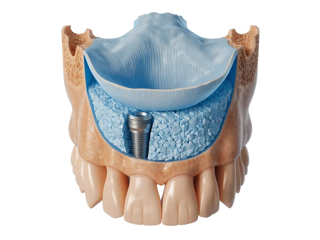
Sinus floor augmentation
This relates specifically to posterior maxillary anatomy. The clinician should assess sinus health and anatomy, residual ridge, implant and prosthetic alternatives, approach, graft category if used, simultaneous or staged placement, possible membrane perforation and the route for medical or specialist input when indicated. A generic “sinus lift” price does not answer those questions.
The procedure name should follow diagnosis, not lead it.
Compare simultaneous and staged care as clinical decisions
Simultaneous augmentation and implant placement combines procedures at one site, while staged augmentation places the graft first and reassesses later implant placement. Neither is automatically faster, safer or better. The choice can depend on defect morphology, ability to place the implant in a restorative position, achieved primary stability, soft-tissue closure, contamination or pathology, augmentation method, patient factors and the clinician's assessment.
A [systematic review of horizontal ridge augmentation before or with implant placement](https://pubmed.ncbi.nlm.nih.gov/24660187/) found different evidence bases and limitations across simultaneous and staged approaches. A [systematic review of simultaneous lateral augmentation](https://pubmed.ncbi.nlm.nih.gov/30675733/) assessed defect resolution across techniques. The studies do not provide a universal defect-size cutoff or calendar.
The written plan should state:
- the intended approach and why;
- which finding permits simultaneous placement;
- which finding triggers staged care;
- what happens if primary stability or position is inadequate;
- whether a provisional tooth changes;
- how the quotation and travel plan change;
- who makes and documents the intraoperative decision.
Consent to a simultaneous plan should include the staged fallback before surgery begins.
Use progression criteria, not a universal healing timetable
Calendar time alone does not prove that a graft is ready for implant placement or that an implant in an augmented site is ready for restoration. Progression can depend on wound healing, absence of unresolved infection or exposure, clinical tissue condition, relevant imaging, graft stability or incorporation as assessed, implant stability, restorative readiness and patient factors. The responsible clinician decides which evidence is needed for the actual procedure.
Avoid fixed promises such as “graft today, implant after exactly X months.” Hospital patient leaflets may describe typical pathways for their own services, but those are not universal clearance dates. Different procedures, sites, donor materials and patients differ.
Ask for decision gates:
| Gate | Evidence owned by the clinician | If not met |
|---|---|---|
| Wound review | Closure, symptoms and tissue condition | Local care, further review or revised plan |
| Graft progression | Site-specific clinical and justified imaging findings | Wait, investigate, revise or abandon augmentation route |
| Implant placement | Restorative position, anatomy and site readiness | Alternative site, further treatment or non-implant option |
| Implant loading | Stability, tissue, restoration and bite criteria | Unloaded healing or interim prosthesis |
| Final restoration | Tissue, impression or scan, component and restorative checks | Delay or redesign |
Travel dates should follow these gates rather than control them.
Describe material sources without product sales
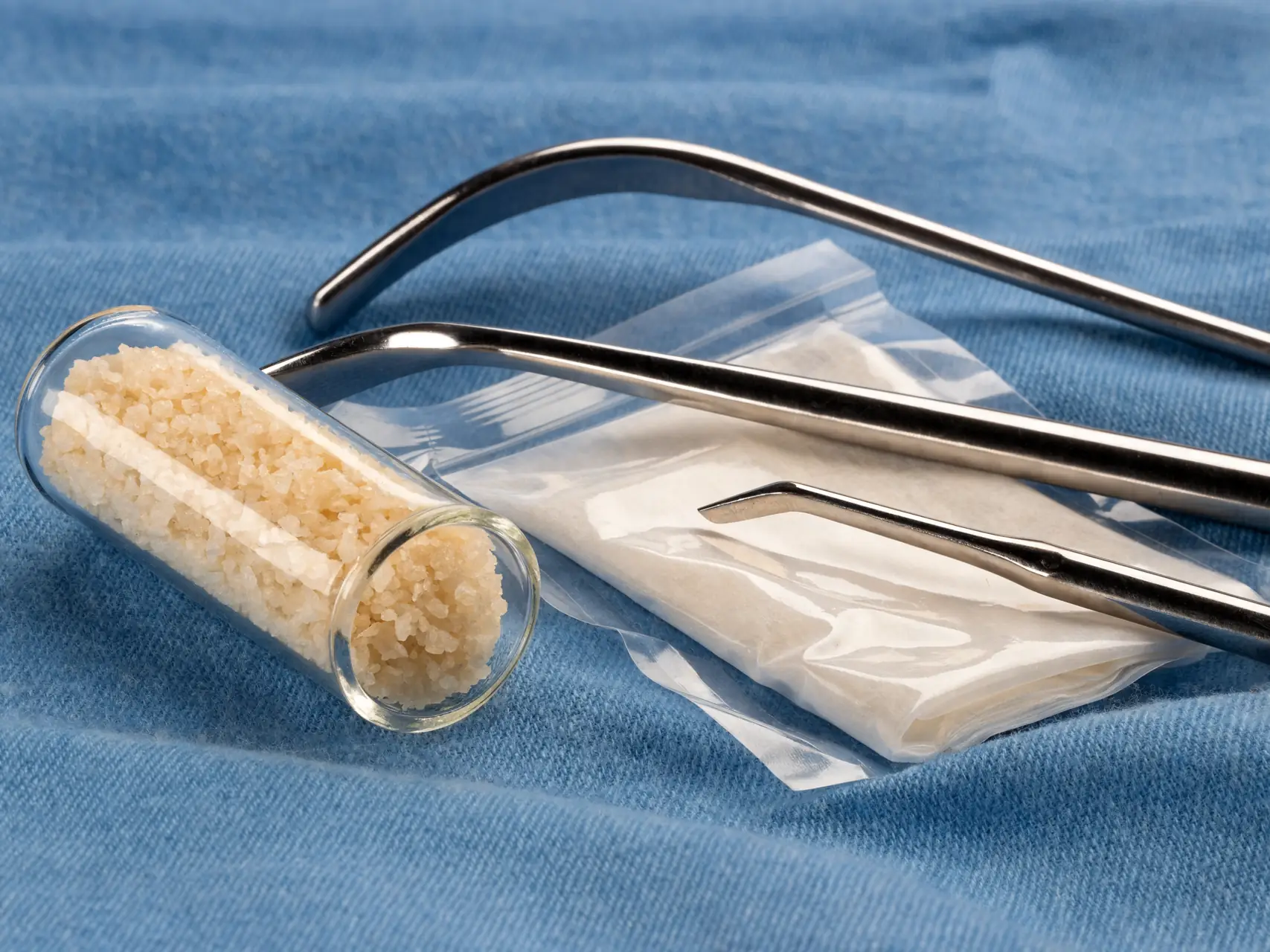
Grafting can involve the patient's own bone, donated human-derived material, animal-derived material, synthetic material or combinations, with membranes, fixation devices or biologic adjuncts in some plans. These are broad categories. Source does not by itself establish indication, safety, effectiveness or superiority.
Autogenous material
Bone from the patient may come from an intraoral or extraoral donor site depending on the required amount and procedure. It creates a second surgical site and additional donor-site risks and recovery considerations. Ask why it is proposed, how much is expected, who performs harvesting and how both sites are managed.
Human-donor material
Ask for the exact product, manufacturer or tissue establishment information where applicable, processing, regulatory status relevant to the place of use, traceability, storage and consent information. Do not rely on the vague phrase “human bone.”
Animal-derived material
Ask for species and tissue source, manufacturer, processing, indications, residual material expectations, religious or ethical concerns and alternatives. “Natural” is not a clinical justification.
Synthetic material
Ask for composition, manufacturer, product indication, handling, resorption or persistence expectations where relevant, and why it suits the defect. “Synthetic” is not automatically free of risk or suitable for every procedure.
The patient should receive enough information to make an informed choice and to carry useful product records home.
Trace membranes, fixation and adjuncts too
The graft particulate or block is not the entire procedure. A plan may include a resorbable or non-resorbable membrane, mesh, pins, tacks, screws, plates, collagen product or another adjunct. Each item can affect removal, exposure management, imaging artefact, future access and traceability.
Request:
- exact device or material name;
- manufacturer;
- lot, batch or unique identifier where assigned;
- source category and composition;
- expiry and storage records as applicable;
- site and quantity recorded clinically;
- fixation map;
- whether removal is intended or conditional;
- patient or custom-device documentation where relevant.
A verbal promise that “premium European materials” are used is not traceability. The surgical record should connect the product actually placed to the patient and site.
Separate material choice from surgical technique
No material can compensate for an unsuitable site, unstable wound, poor closure, unaddressed disease, incorrect implant position or unmaintainable restoration. Similarly, a familiar technique does not make every material interchangeable. The clinician should explain how defect morphology, space maintenance, vascular supply, stability, soft tissue and patient factors inform the combination.
Systematic reviews of grafting studies report varied outcomes and inconsistent definitions. A broad [review of bone preservation and augmentation outcome measures](https://pubmed.ncbi.nlm.nih.gov/35817421/) found major diversity across procedures and reporting. That means a research figure should not be presented as an individual guarantee.
Ask the clinician to name the evidence most relevant to the actual defect and explain its limits. Product brochures and before-and-after photographs are not substitutes for a diagnosis, peer-reviewed evidence and patient-specific consent.
Include sinus-specific boundaries only for posterior maxillary sites
Sinus augmentation is irrelevant to most implant sites and should not be inserted into every bone-graft page as if it were routine. When the posterior maxilla is involved, the clinician should document the restorative position, residual ridge, sinus anatomy and any symptoms or disease that require dental, medical or specialist assessment.
The comparison may include a transcrestal or lateral approach, staged or simultaneous placement, a shorter implant, different prosthesis, alternative site or no implant. The patient should know why one route is preferred and what finding changes it. A [systematic review comparing shorter implants with longer implants and sinus floor elevation](https://pubmed.ncbi.nlm.nih.gov/29980321/) and a [timewise review of randomised studies](https://pubmed.ncbi.nlm.nih.gov/36529571/) support discussing alternatives and uncertainty.
Consent should include site-specific concerns such as membrane perforation, sinus symptoms, infection, graft migration, communication with the sinus, bleeding, swelling, the possible need to stop or alter the procedure, and the route for urgent or specialist care. These are categories for discussion, not a personal risk estimate.
Include GBR and ridge alternatives only for the diagnosed defect
For a horizontal deficiency, clinicians may compare GBR, block graft, ridge expansion or a no-augmentation restorative alternative. For a vertical deficiency, the options and risk profile can be different. A [systematic review of ridge expansion](https://pubmed.ncbi.nlm.nih.gov/38085739/) and a [review comparing horizontal augmentation approaches](https://pubmed.ncbi.nlm.nih.gov/39161939/) show heterogeneous indications and studies; they do not select a method for a patient.
Require a technique-specific explanation:
- What defect shape and objective make this method relevant?
- Which alternative is less invasive or more repairable?
- Is implant placement simultaneous or staged?
- What soft-tissue management is required?
- What exposure, infection, resorption, fracture or donor-site concerns are discussed?
- What evidence determines progression?
- What happens if the achieved contour is less than planned?
Do not consent to a menu of every augmentation technique. Consent to the diagnosed site and its documented branch plan.
Plan the implant restoration before augmenting
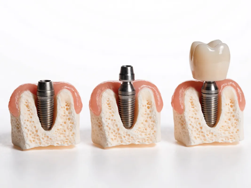
Augmentation should serve an implant position and restoration that are clinically responsible, not simply create radiographic volume. The plan should show the intended crown, bridge or prosthesis; emergence and tissue contour; screw access or retention implications; contacts and bite; hygiene access; and future component service.
If the proposed crown would be over-contoured or difficult to clean even after grafting, reconsider the restorative position, graft objective or treatment option. If an aesthetic contour procedure is proposed, separate the goal of housing an implant from the goal of changing ridge appearance. Each may have different alternatives and consent.
Ask the restorative clinician to approve the augmentation objective before surgery. A surgical record that says “bone added” without the restorative target does not show whether the procedure achieved what the final prosthesis needs.
Identify health and behaviour factors without automatic exclusions
Medical history, medicines, previous radiotherapy, antiresorptive or antiangiogenic medicines, diabetes, immune conditions, bleeding risk, smoking or vaping, periodontal disease, oral hygiene and previous graft or implant history can influence assessment. None should be converted by this page into an automatic approval or refusal.
The patient should provide a current medicine list, allergies and relevant clinician contacts. The dental clinician decides whether medical coordination is needed and who owns it. Do not stop prescribed medicines or change medical care for a commercial itinerary without advice from the responsible prescriber and treating clinicians.
Smoking and hygiene discussions should be specific and non-punitive. Ask how the factor changes the proposed procedure, whether an alternative reduces risk or burden, and what evidence-based support is offered. A consent signature does not transfer every avoidable responsibility to the patient.
Build infection and wound contingencies
The plan should explain how the team responds to wound opening, membrane or mesh exposure, persistent drainage, infection concern, graft mobility, donor-site symptoms, sinus symptoms, bleeding, altered sensation or unexpected pain. A photograph can help communication but cannot rule out every problem.
Before travel, identify:
- the clinical contact and hours;
- the local emergency and urgent dental route;
- which symptoms require same-day local assessment;
- who can remove sutures or assess an exposed device;
- how microbiology, imaging or specialist referral is arranged if needed;
- what records and product details a local clinician receives;
- who authorises intervention and how costs are handled;
- when a return to the original provider is clinically appropriate.
Do not wait for a remote response when breathing, swallowing, systemic illness or rapidly spreading swelling is involved.
Distinguish graft progression from implant progression
A graft can appear clinically satisfactory yet the later implant position or stability may still differ from the plan. An implant can be placed in an augmented site but not yet be ready for loading. A crown can be delayed by tissue, component or restorative findings even after implant review. These are separate progression gates.
Record the owner and evidence for:
- wound and donor-site review;
- graft or reconstructed-site assessment;
- implant-placement decision;
- loading decision;
- provisional restoration;
- final restoration;
- maintenance baseline.
If a provider sells “graft plus implant” as one line item, ask it to unpack these clinical stages and their fallbacks. One payment does not merge the biology.
Use consent as an ongoing process
The [GDC standard on valid consent](https://standards.gdc-uk.org/pages/principle3/principle3) requires GDC registrants to explain relevant options, risks, potential benefits and costs, check understanding, and renew consent if treatment or estimates change. These duties apply to GDC registrants; citing them does not claim that an overseas clinician is GDC regulated. They are a useful UK patient-facing benchmark.
Consent may need distinct stages for extraction, donor-site surgery, grafting or augmentation, membrane and fixation devices, sinus-related surgery, implant placement, sedation or anaesthesia where relevant, loading and restoration. The discussion should cover material source, ethical or religious preferences, alternatives, unresolved findings, the possibility that grafting does not permit the planned implant, and local aftercare.
If the surgical finding changes the material, technique, site, implant timing or price, stop when clinically reasonable, explain the revision and renew consent. A broad form authorising “any necessary graft” does not replace a patient-specific conversation.
Demand an itemised written quotation
The quotation should separate assessment and reports, extraction where proposed, ridge preservation, each graft or augmentation procedure, donor-site surgery, graft material, membrane, mesh or fixation, sedation or anaesthesia where relevant, medicines, implant placement, components, provisional restoration, final restoration, reviews, device removal if intended, maintenance and record release.
It should state:
- what is confirmed and what remains conditional;
- which tooth and site each item belongs to;
- whether implant placement is simultaneous or staged;
- what changes if augmentation is not required or cannot proceed;
- what changes if additional augmentation is proposed;
- whether a donor site creates separate care and costs;
- whether fixation removal is included or conditional;
- how product substitutions are approved and documented;
- deposit, cancellation and refund terms;
- who pays for local assessment, extra travel or changed plans;
- separate suppliers and terms for non-clinical travel.
Compare quotations only after matching scope, material source, stages, records and aftercare. The written quotation controls inclusion; this page makes no package promise.
Collect traceable clinical and material records
The [GDC standard on patient information and records](https://standards.gdc-uk.org/pages/principle4/principle4) includes complete clinical records, radiographs, photographs, models, consent documents, laboratory prescriptions and statements of conformity where available. This is a direct duty for GDC registrants and a useful records benchmark, not a claim about another jurisdiction.
Request, as relevant:
- tooth prognosis and site diagnosis;
- medical history and coordination notes;
- images and reports in usable formats;
- restorative and implant position plan;
- augmentation objective and technique;
- donor-site and recipient-site surgical record;
- graft, membrane, mesh, fixation and adjunct product traceability;
- lot, batch or identifier where assigned;
- implant system and component details;
- medicines and discharge information;
- wound, graft and implant progression records;
- provisional and final restoration records;
- named providers, clinicians and laboratories;
- maintenance, urgent-care and complaint routes.
Check that records identify what was actually placed, not only what appeared in the pre-treatment quote.
Plan travel around clinical gates
The [GDC guide to dental treatment abroad](https://www.gdc-uk.org/docs/default-source/information-standards-and-guidance/going-abroad-for-dental-treatment/going-abroad-for-your-dental-cared3d463b7cee0446882bf877483299dd4.pdf?sfvrsn=c67e2b57_7) advises patients to consider providers, regulation, qualifications, assessment, risks, costs, records, aftercare and complaint routes. A home dentist may help identify record or maintenance gaps before travel.
Use flexible arrangements around examination, surgery, wound review, graft progression, implant placement, loading and restoration. Ask what can change after direct assessment, what local care is needed between stages and how a postponed gate affects travel and payments. Procedure-specific advice about flying, lifting, food, activity, medicines and assistance should come after the actual treatment.
Non-clinical transfers and accommodation should be separately identified. They do not establish clinical readiness. The dental treatment timeline guide helps separate decision gates from calendar marketing, and the returning home after dental tourism guide supports handover planning.
Arrange local aftercare before grafting
Ask a home dentist or suitable local provider before travel whether they will assess healing and what records they require. A local clinician decides what they can safely manage and may not remove unfamiliar fixation, interpret another provider's intended progression or assume responsibility for an overseas plan.
The handover should identify:
- routine wound review;
- suture and donor-site care where applicable;
- symptoms requiring earlier assessment;
- access to images, reports and product details;
- responsibility for graft and implant progression decisions;
- periodontal and hygiene maintenance;
- urgent escalation and specialist referral;
- payment and communication boundaries;
- what requires return to the original provider.
Remote review is useful for communication, not a substitute for examination when clinical findings matter. Use the dental implant service guide for the broader implant assessment context and the not-enough-bone planning guide for the patient-level differential.
Know urgent and emergency red flags
Seek prompt professional assessment for severe or worsening pain, increasing swelling, fever or marked illness, persistent discharge, unpleasant taste with other symptoms, uncontrolled bleeding, wound opening, visible or mobile membrane or fixation, graft mobility, new or changing altered sensation, donor-site problems, sinus symptoms, or inability to maintain food and fluid intake. The treating clinician should personalise discharge thresholds.
Difficulty breathing or swallowing, rapidly spreading swelling, collapse, major facial bleeding or another life-threatening concern requires the current local emergency route. Do not wait for a coordinator or a return flight.
Do not push material back into a wound, remove fixation, repeatedly probe the site or self-prescribe leftover antibiotics. Keep detached material or devices when safe and bring the treatment and product records to the assessing clinician.
Red flags in a graft-and-implant proposal
Pause when a proposal:
- recommends extraction without a tooth-specific prognosis;
- says the person “has no bone” without identifying the site and restorative need;
- treats a graft as mandatory without a reasonable no-graft comparison;
- promises that grafting will make implant treatment possible;
- prescribes CBCT or advanced imaging without a defined question and responsible interpretation;
- offers the same graft technique for every defect;
- promises simultaneous implant placement before clinical gates are known;
- gives a fixed healing or travel timetable as biological clearance;
- sells donor or synthetic material as universally superior;
- cannot identify source, manufacturer, lot or fixation records;
- ignores soft tissue, restoration, bite or hygiene access;
- has no fallback for exposure, infection or insufficient progression;
- hides donor-site, membrane, fixation, removal or local aftercare costs;
- cannot name the legal provider and responsible clinicians;
- makes flights or hotel dates control clinical progression;
- supplies no transferable records or local urgent-care route.
One missing answer may be resolved. Repeated pressure, vague responsibility and unsupported certainty justify another opinion.
A site-specific decision worksheet
| Decision gate | Evidence required | Named owner | Fallback |
|---|---|---|---|
| Preserve or extract | Tooth-specific prognosis and alternatives | Examining clinician | Treatment, monitoring, referral or revised plan |
| Define deficiency | Restorative position, anatomy, tissue and site map | Surgical and restorative clinicians | Different restoration or no implant |
| Graft or alternative | Technique-versus-no-graft comparison | Named treating clinician | Shorter/different implant, prosthesis or no treatment where relevant |
| Material and device | Source, product, traceability and consent | Clinician and legal provider | Approved alternative or stop |
| Simultaneous or staged | Site morphology, position, stability and closure | Surgical clinician | Staged graft, unloaded healing or alternative |
| Graft progression | Clinical and justified imaging evidence | Treating clinician | Wait, investigate, revise or abandon route |
| Implant progression | Placement and loading criteria | Implant clinician | Unloaded healing or alternative restoration |
| Handover | Records, local provider and urgent route | Treating and home clinicians | Referral or return assessment |
The plan is not ready while a critical gate has no evidence, owner or contingency.
Questions to ask the named clinician
- Can each tooth proposed for extraction be preserved?
- Which exact implant site and restorative position lack bone?
- Is the deficiency horizontal, vertical, socket-related, sinus-related or combined?
- Which clinical and imaging records support that conclusion?
- What remains unknown until direct examination or surgery?
- What no-graft alternatives apply to this site?
- Why is this augmentation category relevant?
- Why is implant placement intended simultaneously or later?
- Which finding changes the plan from simultaneous to staged?
- Which graft source and product are proposed, and why?
- Which membrane, mesh, fixation or adjunct is planned?
- What donor-site risks and care apply if my own bone is used?
- How will products and devices be traced in my records?
- Which evidence permits progression to implant placement?
- What happens if the graft is exposed, infected or insufficient?
- How does the graft support the planned crown or prosthesis?
- What is itemised, conditional and excluded in the quotation?
- Who owns routine and urgent aftercare when I am home?
- Which records will be supplied to my local dentist?
- Which symptoms require prompt local or emergency assessment?
Sources and evidence limits
This guide uses official patient information, professional standards and peer-reviewed systematic reviews. It does not turn research averages into an individual prediction. Augmentation studies vary in defect, site, technique, material, timing, implant, restoration, outcome definition and follow-up. Some report radiographic gain, some implant survival, some complications and relatively few patient-reported outcomes. A favourable measurement in one domain does not prove that a graft, implant and restoration will all meet a patient's goals.
Key sources include:
- [Cambridge University Hospitals: Bone grafting for dental implants](https://www.cuh.nhs.uk/patient-information/bone-grafting-for-dental-implants/) and [Dental implants in restorative dentistry](https://www.cuh.nhs.uk/patient-information/dental-implants-in-restorative-dentistry/).
- [Guy's and St Thomas' NHS Foundation Trust: Bone grafting for dental implants](https://www.guysandstthomas.nhs.uk/health-information/bone-grafting-dental-implants).
- [GDC Principle 3: Obtain valid consent](https://standards.gdc-uk.org/pages/principle3/principle3), [GDC Principle 4: Maintain and protect patient information](https://standards.gdc-uk.org/pages/principle4/principle4), and [GDC: Going abroad for dental care](https://www.gdc-uk.org/docs/default-source/information-standards-and-guidance/going-abroad-for-dental-treatment/going-abroad-for-your-dental-cared3d463b7cee0446882bf877483299dd4.pdf?sfvrsn=c67e2b57_7).
- Systematic reviews of [horizontal augmentation before or with implant placement](https://pubmed.ncbi.nlm.nih.gov/24660187/), [simultaneous lateral augmentation](https://pubmed.ncbi.nlm.nih.gov/30675733/), [augmentation outcomes and measures](https://pubmed.ncbi.nlm.nih.gov/35817421/), [ridge expansion](https://pubmed.ncbi.nlm.nih.gov/38085739/) and [comparative horizontal augmentation approaches](https://pubmed.ncbi.nlm.nih.gov/39161939/).
- Systematic reviews of [immediate placement versus ridge preservation](https://pubmed.ncbi.nlm.nih.gov/34100157/) and [ridge preservation versus early placement](https://pubmed.ncbi.nlm.nih.gov/38850092/).
- Reviews comparing [shorter implants with longer implants and sinus elevation](https://pubmed.ncbi.nlm.nih.gov/29980321/), [randomised studies across time](https://pubmed.ncbi.nlm.nih.gov/36529571/), [longer follow-up](https://pubmed.ncbi.nlm.nih.gov/36836403/) and [umbrella-review evidence quality](https://pubmed.ncbi.nlm.nih.gov/32951871/).
The decision is ready only when the named clinicians can define the deficiency at the intended implant position, justify augmentation against alternatives, explain material and staging choices, document the progression and contingency gates, and provide a record and aftercare plan that remains usable after the patient returns home.