A missing front tooth is visible, emotionally important and often urgent from the patient’s point of view. That urgency does not create an automatic indication for an implant. An incisor or canine may be restorable, temporarily replaceable, suitable for orthodontic space closure, replaceable with an adhesive bridge or conventional bridge, or deliberately monitored while growth, trauma or disease is managed. An implant-supported crown is one option among several, and its surgical survival does not guarantee that the gum line, papilla, shade, contour, speech or smile will match the neighbouring teeth.
The “aesthetic zone” is not defined only by the tooth number. It is the part of the mouth visible in the patient’s natural smile and speech. A maxillary central incisor is commonly prominent, but a lateral incisor, canine or lower front tooth may also be highly visible depending on lip movement and smile line. A good decision therefore begins with the person’s face, growth, tooth history, biology and expectations—not a stock implant protocol.
This is an evidence-led decision guide for adults and families researching a front tooth dental implant, including treatment after trauma, infection or developmental absence and treatment considered abroad. It is not an individual diagnosis, prescription, quotation, timetable or appearance promise. Photographs and scans can organise questions but cannot determine tooth restorability, implant suitability or consent remotely. The named clinicians who examine the patient, interpret the required records and accept responsibility for each stage must make and document those decisions.
A front-tooth gap is a diagnosis problem before it is an implant problem
First identify why the tooth looks compromised or is absent. A dark, mobile, fractured or painful tooth is not necessarily non-restorable. A visible gap may result from trauma, congenital absence, extraction, periodontal disease, failed root-canal treatment, root resorption, a retained primary tooth, an impacted tooth, orthodontic movement or loss of an older bridge. Each cause creates a different sequence and different options.
Ask for a written problem list that separates:
- the condition and prognosis of the tooth, if it remains;
- pulp, root, periodontal and surrounding bone findings;
- trauma history and prior emergency or restorative care;
- infection, swelling, drainage or apical disease;
- the position and health of neighbouring teeth and their roots;
- the available space and whether orthodontic treatment has moved or retained it;
- jaw growth and continuing eruption where age or development is relevant;
- ridge contour, hard tissue and soft tissue at the proposed site;
- smile line, lip movement, gingival display and the matching tooth;
- bite, guidance, parafunction, speech and restorative space;
- the patient’s priorities, tolerance for surgery and maintenance capacity;
- every reasonable retention, replacement and no-treatment alternative.
If a sales discussion starts with a brand, same-day tooth, package or before-and-after photograph before these questions are answered, it has skipped the diagnosis.
Urgent trauma and infection come before elective replacement
A recent front-tooth injury may require urgent local assessment. The International Association of Dental Traumatology guidelines cover fractures and luxations of permanent teeth and emphasise proper diagnosis, radiographic assessment, photographic documentation, pulp testing, stabilisation where indicated, patient instructions and follow-up. A tooth that looks displaced, fractured or “lost” should not be converted immediately into a travel implant enquiry. Timely trauma care may preserve the tooth, surrounding tissues and future options.
If a permanent tooth has been completely knocked out, follow current emergency dental guidance and seek immediate local professional help; do not wait for an overseas quote. The correct first aid depends on the tooth and circumstances. Primary teeth are managed differently from permanent teeth. Where there is head injury, loss of consciousness, facial fracture, uncontrolled bleeding, breathing difficulty or another medical emergency, emergency services take priority.
Infection also needs diagnosis. Rapidly increasing facial or neck swelling, difficulty breathing or swallowing, uncontrolled bleeding or collapse requires emergency assessment. Fever, discharge, worsening pain, a spreading swelling, persistent altered sensation or inability to eat and drink warrants prompt professional advice. Antibiotics do not replace examination, drainage or definitive care, and there is no universal patient-facing antibiotic regimen for an anterior implant site.
After trauma or infection, the treatment sequence may remain uncertain while pulp, periodontal ligament, root, bone or soft tissues are monitored. That uncertainty is not a failure. It is a reason to protect options with an appropriate interim restoration rather than rush into extraction or implant placement.
Preserve a restorable front tooth before planning replacement
Extraction is irreversible. The American Association of Endodontists position statement says retention should be the first consideration for a compromised natural tooth and warns against treating implants as a cure-all. It also recognises that implants can be useful when a tooth is missing or cannot be saved. The responsible decision is therefore not “tooth versus implant” as competing products; it is whether retention is reasonable for this tooth and this patient.
A front tooth may need restorative, endodontic, periodontal, orthodontic or trauma-informed assessment. Possible retention categories include repair of a fracture, reattachment of a fragment, direct composite restoration, root-canal treatment or retreatment, management of a resorptive defect, periodontal treatment, splinting in an appropriate trauma pathway, orthodontic repositioning, a crown or veneer where structurally justified, or observation. None can be recommended from this page, and not every tooth can be retained.
Ask the clinician to record:
- the exact diagnosis and source of symptoms;
- remaining sound tooth structure and fracture extent;
- pulp and apical status;
- root form, resorption, crack or fracture concerns;
- periodontal attachment, mobility and bone support;
- the condition of existing restorations or root-canal treatment;
- the likely burden, uncertainty and maintenance of retention;
- why extraction is proposed if retention is rejected;
- whether an appropriate endodontic, restorative, periodontal or trauma opinion is needed.
A second opinion is proportionate before removing a visible tooth when the diagnosis is uncertain, the tooth has strategic value, or the replacement plan involves advanced augmentation. Once extraction occurs, the option to retain that tooth is gone.
Trauma records protect both treatment and future choices
Trauma can affect more than the visibly damaged crown. Keep the date and mechanism of injury, emergency notes, photographs, radiographs, pulp test results, splint details, medicines and follow-up findings. Account for fragments and soft-tissue injuries where clinically relevant. A history of avulsion, intrusion, luxation, root fracture or alveolar fracture changes the questions about the tooth, ridge, adjacent roots and growth.
Repeated pulp testing and imaging may be required according to the injury and clinician’s judgment. An early negative sensibility response does not always establish permanent pulp necrosis after trauma. Conversely, a tooth that looks comfortable can develop later complications. A patient should not be told that a single remote image conclusively proves extraction.
If the injury occurred during growth, the team may need to preserve ridge development while delaying an implant decision. Interim adhesive, removable, orthodontic or composite strategies can maintain appearance or space. In selected ankylosis situations, specialised options may be discussed to manage the ridge, but they require a growth- and trauma-informed clinician and cannot be selected by a generic timeline.
Growth matters even when a teenager looks fully grown
An osseointegrated implant behaves more like an ankylosed unit than an erupting natural tooth. Adjacent teeth and jaws can continue to erupt and develop, leaving an implant crown in relative infra-position or with a changed gum line and contact relationship. This is particularly important in the anterior maxilla, where small positional changes may be visible.
The current American Academy of Pediatric Dentistry best-practice document on periodontal conditions says conservative management is generally indicated for growing patients with missing teeth and that assessment of growth and development is central to implant decisions. It warns that chronological age alone is not a reliable indicator of completed growth. Systematic review evidence also reports infra-position as a long-term concern and acknowledges limited certainty about who will be affected.
Do not use a birthday as the only clearance for an implant. A multidisciplinary assessment may consider serial growth records, facial and dental development, eruption, orthodontic status, skeletal maturation and the consequences of waiting. Exceptional clinical circumstances exist, but they should be explained and monitored by clinicians competent in paediatric, orthodontic, restorative and surgical care.
While growth continues, options may include:
- retaining and monitoring a useful primary tooth where appropriate;
- orthodontic space closure or redistribution;
- a resin-bonded provisional or definitive bridge where suitable;
- a removable prosthesis;
- composite reshaping of neighbouring teeth after orthodontic movement;
- a temporary tooth attached to an orthodontic retainer;
- monitoring with a defined future review point.
These are not inferior waiting-room solutions. They can preserve tissue and flexibility until a definitive decision is safer. The patient and family should understand repairs, replacement, hygiene, appearance and review needs.
Space and root position can make or break an implant plan
The visible width of a gap does not prove that an implant can be placed safely or restored naturally. Adjacent roots may converge beneath apparently adequate crown space. Teeth may have drifted, rotated or tipped. An unerupted or impacted tooth may occupy the path. The space may be too narrow for a proportional crown, or too wide for a natural contact and papilla. The opposing bite may leave inadequate restorative clearance.
Orthodontic treatment can close a space completely or create and maintain the position needed for a later replacement. The British Orthodontic Society’s missing-teeth information describes braces as a way to close gaps or create appropriate space for a future bridge, denture or implant. That decision involves facial profile, bite, tooth shapes, root positions, midlines, growth, treatment burden and the restorative plan.
An implant should not be placed merely because braces have created a visible gap. The restorative and surgical teams should confirm:
- the intended final crown width, length and position;
- root separation along the full proposed implant path;
- the position of the midline and contact points;
- the gingival margins and bone levels of neighbouring teeth;
- sufficient room for components and cleanable contours;
- the retention plan after orthodontics;
- whether composite reshaping or another restorative addition is expected;
- what happens if teeth move before surgery or while the implant heals.
Orthodontic retainers and temporary teeth can affect soft tissue and space. The team should agree who adjusts them before and after surgery. A patient travelling for implant placement should have current orthodontic records and a local plan to maintain the space.
Examine the smile, not just the empty socket
A front-tooth restoration is seen within a moving face. Clinical photographs should record a natural smile, full smile, rest position, speech-related tooth display, profile and close views with a calibrated shade reference where appropriate. The assessment may include lip mobility, gingival display, smile arc, dental midline, tooth proportions, incisal edge position, symmetry and the relationship to the lower lip.
A high smile line may reveal tissue differences that are hidden in a low smile. A matching central incisor creates different symmetry demands from a lateral incisor. A canine contributes to arch form and function as well as appearance. Lower incisors may be less visible in a youthful posed smile but more visible in speech or with age. The patient should demonstrate what matters to them rather than accept an assumed beauty template.
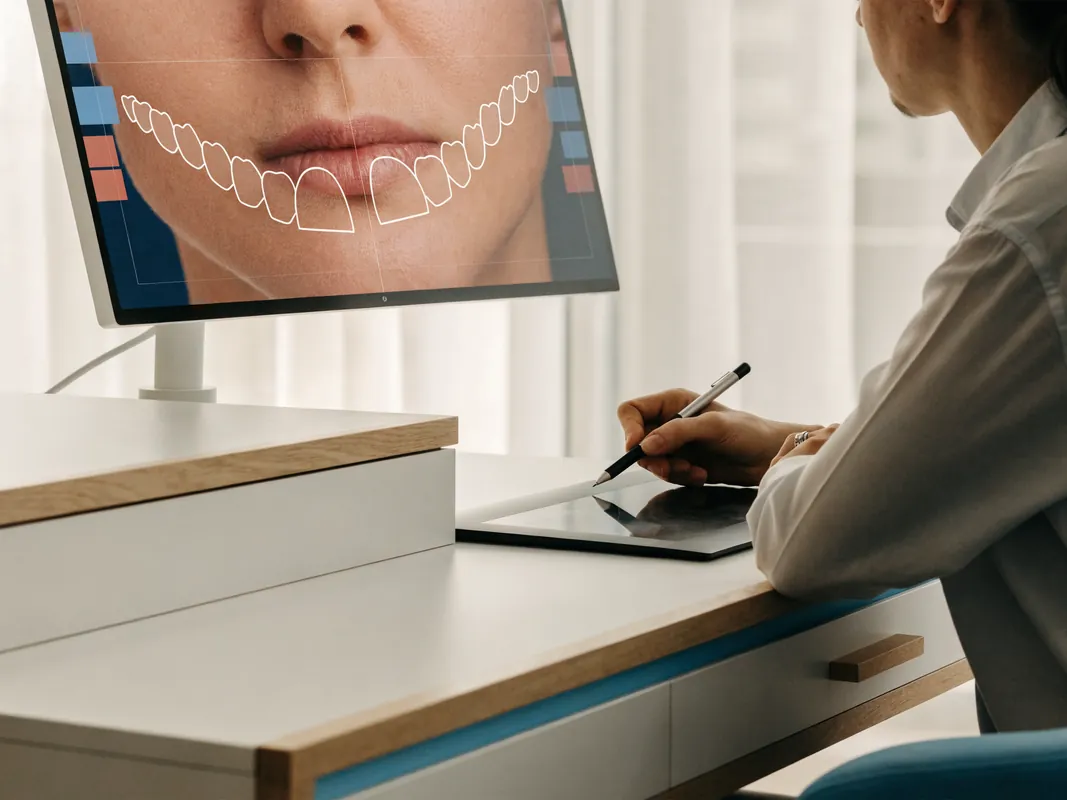
Photographs are records and communication tools. They do not predict a result. Lighting, lens, distance, angle, retraction, dehydration, editing and screen calibration can change apparent colour and proportions. Before-and-after images from another patient do not show this patient’s biology, smile or future tissue behaviour. Any simulation or digital smile design must be labelled as a planning aid, not a guarantee.
Gingival phenotype, tissue contour and inflammation require separate assessment
The gums around a front implant influence colour, contour, papilla and the visibility of the restorative transition. Clinicians may assess tissue thickness or phenotype, keratinised tissue, recession, scarring, vestibular depth, mucosal mobility, periodontal health and the shape of the ridge. Terms such as “thin biotype” are not a complete diagnosis and do not automatically prescribe a connective-tissue graft.
Inflammation must be controlled. Plaque, bleeding, periodontal pockets, active disease or a provisional that traps debris can undermine both health and appearance. The EFP S3 guideline on peri-implant diseases supports periodontal risk control, prosthetic designs that permit hygiene and ongoing supportive care. A photo-ready emergence profile that the patient cannot clean is not a successful long-term design.
Soft-tissue augmentation may be considered in selected cases. Systematic review evidence evaluates connective-tissue grafting around immediate implants, but it does not make grafting mandatory for every anterior site or promise stable symmetry. Donor-site discomfort, bleeding, healing, texture, colour, scar and incomplete correction belong in consent. Alternative soft-tissue, prosthetic or no-graft plans should be discussed where relevant.
Bone, socket walls and adjacent teeth shape the aesthetic risk
Hard-tissue assessment should identify ridge width, height, contour, facial and palatal or lingual walls, defects, infection, root positions and relevant anatomical boundaries. The bone on adjacent natural teeth matters because it helps support the papilla between tooth and implant. A site with intact neighbouring attachment presents a different aesthetic question from two adjacent missing teeth, periodontal bone loss or a vertical defect.
Extraction sockets remodel even when an implant is placed. Immediate placement does not “freeze” the ridge or guarantee preservation of the gum line. Bone grafting or contour augmentation may be discussed, simultaneously or in stages, but each adds material, wound and contingency questions. The intended implant and crown position should guide the decision; filling a defect without a prosthetic plan is not enough.
Avoid universal millimetre rules on a public page. Site dimensions, implant design, soft tissues, adjacent roots, prosthesis and surgical technique interact. The clinician should show the defect in relation to the intended crown and explain what finding would make the preferred plan change.
Papilla and gum-line appearance remain uncertain
The papilla is the tissue peak between teeth. Patients often fear a dark embrasure or “black triangle.” A systematic review on papilla height around single-tooth implants found limited evidence that the relationship between the interproximal contact and crestal bone matters, while the periodontal attachment of the neighbouring tooth is important. Osteology Foundation consensus work also highlights that tissue fill between two adjacent implants is less straightforward and that one universal optimal spacing rule could not be defined from the evidence.
This means a clinician can assess risk and design the restoration, but cannot promise complete papilla fill. The neighbouring tooth’s attachment, the defect, number and position of implants, tissue, contact shape, provisional contour, inflammation, healing and time all contribute. A photograph of a successful case does not remove this uncertainty.
Consent should address possible recession, asymmetry, dark triangles, elongated contact areas, a grey hue through tissue, altered crown proportions, a visible transition or the need for pink restorative material. These are not merely cosmetic footnotes. They may determine whether an implant, adhesive bridge, orthodontic option or removable prosthesis best fits the patient’s priorities.
Imaging must be justified and interpreted in clinical context
Current ADA and American Academy of Oral and Maxillofacial Radiology recommendations place history and clinical examination before imaging selection. Intraoral or panoramic images can answer some questions. CBCT may be justified for presurgical implant planning because it shows three-dimensional relationships, but the field of view and exposure should match the clinical need and the entire acquired volume must be interpreted by someone competent.
CBCT cannot replace tooth testing, periodontal probing, tissue examination, trauma assessment, occlusal analysis or a medical history. A remote DICOM file may be useful, but date, quality, artefact, field of view and lack of examination limit conclusions. The patient should receive a documented interpretation and copies suitable for future care. Repeat imaging should have a clinical reason rather than being required automatically for marketing or administration.
Build a complete medical, dental and functional baseline
An implant decision includes the whole patient. Record medical conditions, medicines, allergies, previous adverse events, diabetes and relevant control information, bleeding risk, immune conditions, cancer treatment, radiotherapy, antiresorptive or antiangiogenic medicines, steroid exposure, smoking or nicotine use, alcohol or drug use, pregnancy possibility and anaesthesia history. These factors do not create a simple public yes-or-no rule; they identify clinician-owned risk and liaison questions.
Dental history should include caries, periodontal disease, previous trauma, endodontic care, orthodontics, missing teeth, implants, parafunction, dry mouth, maintenance and ability to clean. The functional examination may consider overbite, overjet, anterior guidance, excursive contacts, tooth wear, clenching or grinding, opposing teeth, mobility and restorative space. A canine replacement may carry different functional demands from a lateral incisor.
Pain, clicking or jaw-muscle symptoms need their own baseline rather than being attributed automatically to the missing tooth. An implant crown cannot be promised to cure jaw pain. The restoration should be planned within an individual occlusal scheme and reviewed for contacts after fitting and over time.
Compare all reasonable front-tooth replacement options
An informed plan should include retention where possible, no immediate replacement, and realistic non-implant replacements. The alternatives are not token sentences before selling an implant. They should be compared for tooth preparation, surgery, appearance, function, repair, expected maintenance, uncertainty, cost and local access.
No immediate replacement
Deferring replacement may be reasonable while disease or trauma is monitored, growth continues, orthodontic planning is completed, the patient considers options or a temporary arrangement is acceptable. The space, neighbouring teeth, appearance, speech and psychosocial effect should be reviewed. “Do nothing now” is not the same as abandoning care; it requires a defined review and space-management plan.
Direct composite repair or tooth reshaping
If a tooth remains, direct composite may repair a fracture, replace missing contour, mask a limited defect or reshape neighbouring teeth after orthodontic space closure. It preserves options and can be repaired, but it can stain, wear, chip or require maintenance. Composite cannot resolve every structural, periodontal or positional problem. The treating dentist must decide whether the tooth and bite support it.
Orthodontic space closure or redistribution
Orthodontic movement may close a lateral-incisor space, redistribute spacing, correct root position or prepare a site for another restoration. It can avoid an implant in some cases but changes tooth positions, shape relationships and occlusion. Composite reshaping, whitening or gingival adjustment may later be considered. Retention is part of the plan. The orthodontist and restorative clinician should agree on the target before movement begins.
Resin-bonded or adhesive bridge
A resin-bonded bridge commonly uses a wing bonded to a neighbouring tooth to support a replacement tooth. It can preserve more tooth tissue than a conventional bridge and avoids implant surgery. Suitability depends on enamel, abutment condition, space, bite, moisture control, material, design and patient habits. Debonding can occur, which may be repairable but still creates inconvenience and cost.
Systematic reviews support including anterior resin-bonded bridges in the option discussion, while also showing variation in designs, materials and evidence. A single-retainer design may be considered in appropriate cases, but a public page cannot prescribe it. The patient should understand which tooth is used, how much preparation is planned, whether the bridge is intended as interim or definitive and how a debond would be managed.
Conventional tooth-supported bridge
A conventional bridge may be considered when neighbouring teeth are already heavily restored or would benefit from crowns, or when other clinical factors support it. It usually requires more preparation of abutment teeth than an adhesive design. Risks can include biological and technical complications involving the supporting teeth, margins, pulp, caries, periodontal tissues or framework. The comparison should consider the actual condition of the adjacent teeth rather than treating bridge preparation as automatically wrong.
Removable prosthesis
A removable partial denture, temporary “flipper” or other removable design can replace one or several front teeth. It may be useful during growth, healing, staged treatment or when surgery is not chosen. It can also affect speech, comfort, retention, plaque and soft-tissue pressure. A removable provisional over a graft or implant site must be designed and adjusted so it does not harm the surgical plan.
Implant-supported crown or bridge
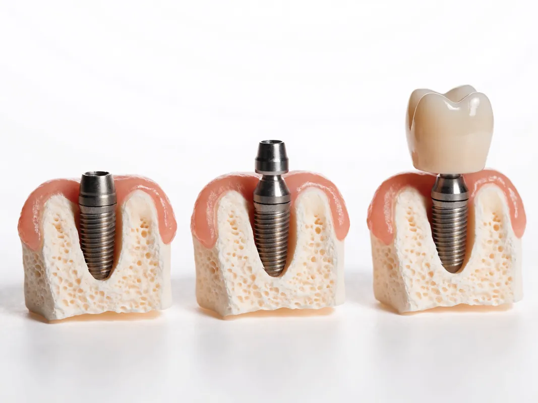
An implant can avoid preparing neighbouring teeth and may support a single crown or selected multi-unit design. It adds surgery, anatomical constraints, component dependence and lifelong implant maintenance. For adjacent missing front teeth, implant number and distribution require special attention to papilla, restorative contours and cleanability. One implant supporting a short cantilever, two implants, an adhesive bridge or another solution are case-specific categories, not universal hierarchies.
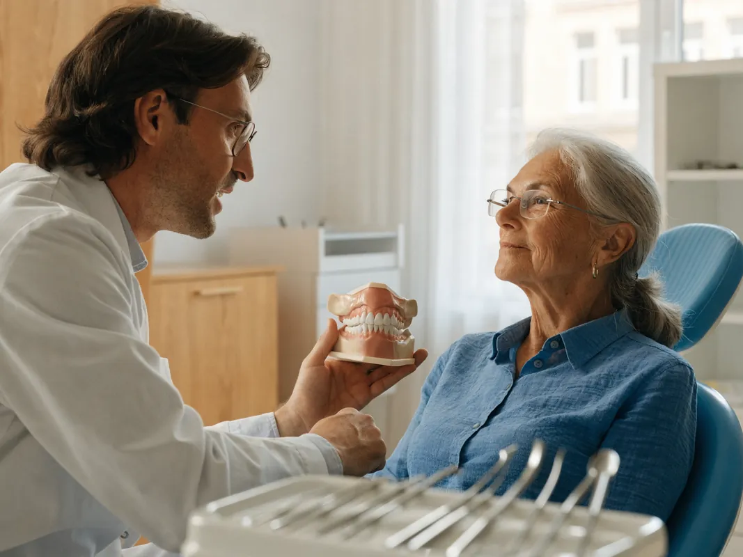
The comparison should be personalised. A healthy adolescent with trauma, an adult with intact adjacent teeth, an adult with crowned neighbours and a patient with periodontal loss may reasonably choose different paths.
Plan from the final crown backwards
Anterior implant surgery should be prosthesis-led. Define the intended crown edge, facial contour, contact points, emergence, incisal position, shade, screw-access or cementation considerations and hygiene access before selecting the fixture position. The question is not simply where bone exists. It is whether a maintainable restoration can emerge in the right place without creating unacceptable biological, mechanical or visual compromise.
A diagnostic set-up, wax-up, digital design or provisional may help test the plan. It should show:
- crown width, length and symmetry;
- incisal edge and speech relationship;
- gingival margin and contact design;
- emergence contour and space for tissue;
- relationship to adjacent roots and planned implant path;
- restorative thickness and abutment space;
- access for cleaning and future component work;
- compromises if the ridge or gum cannot be reconstructed fully.
Digital planning and surgical guides can help transfer a plan but do not remove error or intraoperative judgment. Guide support, fit, mouth opening, tissue movement, manufacturing tolerance and surgical findings may change execution. Consent should explain whether the approach is guided, partially guided or freehand and what fallback applies if the simulated position cannot be used.
Extraction timing and implant timing are different decisions
Several events are often collapsed into “same-day implant.” Keep them separate:
- the decision that a tooth is non-restorable;
- extraction timing and technique;
- management of infection or trauma;
- socket or ridge-preservation measures;
- implant placement timing;
- augmentation timing;
- loading timing;
- provisional restoration;
- final restoration.
An implant placed at extraction is called immediate placement. It is not automatically an immediate-load implant and does not prove that a fixed temporary or final tooth can be delivered that day. Delayed or early placement categories separate extraction and implant surgery for clinical reasons. No category is universally best for the aesthetic zone.
The 2023 ITI consensus and its supporting systematic review consider immediately placed and immediately loaded single implants a clinically viable option in selected maxillary aesthetic-zone cases while acknowledging surgical, technical and biological complications. That evidence does not justify an “almost always” promise. Published studies use selection criteria and professional protocols; the named clinician must determine whether those conditions relate to the individual.
Immediate placement is not a tissue-preservation guarantee
Immediate placement may reduce the number of surgical episodes in a selected site, but it does not stop socket remodelling or guarantee papilla and midfacial gum stability. Site anatomy, facial bone, soft tissue, infection, extraction damage, implant position, gap management, stability and provisional design matter. A traumatic extraction or poorly positioned fixture cannot be rescued by the word immediate.
The preoperative plan should identify the criteria for immediate placement and the fallback if they are not met. If the facial socket wall is damaged, infection is more extensive, the root position is unfavourable, stability is inadequate or the correct restorative position cannot be achieved, options may include socket management and later placement, augmentation, another replacement or stopping. The patient should consent to this branch before the tooth is removed.
Never let a non-refundable flight or package turn immediate placement into an obligation. If examination or surgery changes the diagnosis, the patient must have time to understand the revised plan and costs.
Bone augmentation is conditional and prosthetically justified
Socket management, local contour grafting, guided bone regeneration or staged ridge augmentation may be discussed where tissue conditions and the intended restoration support them. These procedures differ in material, wound, donor-site, fixation and complication burden. They should not be described as routine additions to every front-tooth implant.
Ask:
- What defect is being treated?
- Is the purpose fixture support, contour, prosthetic position or tissue appearance?
- Is augmentation simultaneous with placement or staged?
- Which graft, membrane, fixation or adjunct is proposed?
- What is the tissue source and exact product where known?
- What allergy, ethical, religious or preference issues matter?
- What risks include exposure, infection, incomplete gain or further surgery?
- How will a removable temporary avoid pressure on the site?
- What evidence will be used before progressing to the next stage?
- What happens if the intended contour is not achieved?
No graft creates a guaranteed natural gum line. The restorative plan may need a longer crown, altered contact, pink ceramic or composite, a removable option or another compromise. Those possibilities belong in the original consent, not only after disappointment.
Soft-tissue augmentation is not automatic
Connective-tissue or other soft-tissue procedures may be discussed to alter thickness or contour in selected cases. The evidence suggests potential effects in certain protocols, but aesthetic findings and methods vary. A graft cannot recreate a periodontal ligament around an implant or guarantee the papilla, shade masking or lifelong margin stability.
The plan should explain the donor site, technique, expected recovery, risks, alternatives and uncertainty. Possible burdens include pain, bleeding, swelling, wound problems, altered texture or colour, scarring and incomplete correction. If tissue is already adequate and cleanable, a graft may add burden without a clear patient-valued benefit. The decision belongs to the named clinician and patient after examination.
One missing tooth and several adjacent teeth are different problems
A single missing front tooth bounded by healthy natural teeth can use adjacent tooth attachment and contacts to help shape the papilla. Two adjacent missing incisors remove that tooth-implant relationship in the centre, creating a different tissue and prosthetic problem. Several missing teeth may also reflect trauma, congenital absence, periodontal loss or previous surgery, each with different ridge form.
For two adjacent sites, options may include two implants, one implant supporting a two-unit design in selected circumstances, an adhesive or conventional bridge, orthodontic redistribution or a removable prosthesis. Evidence for cantilever designs and adjacent implants is case-specific and does not establish a universal winner. Space, roots, occlusion, tissue, materials, component access and repair matter.
Avoid fixed statements such as “one implant is always better for papilla” or “one implant per tooth is ideal.” The restorative team should demonstrate the proposed contacts, contours, support and hygiene access and explain what tissue fill cannot be promised.
Placement, loading, provisional and final stages must stay distinct
Clear terminology protects consent:
- placement is surgical insertion of the fixture;
- immediate placement describes timing relative to extraction;
- loading means applying prosthetic load according to a defined protocol;
- a provisional restoration is an interim tooth used during assessment or healing;
- a final restoration is selected and fitted after the relevant clinical gates;
- integration is a biological process assessed over time, not proved by a same-day photograph.
An immediately placed implant may be unloaded. A provisional may be removable or bonded to adjacent teeth rather than attached to the implant. A fixed provisional may be shaped to support communication and tissue development but may carry contact or dietary restrictions. It is not the final crown and may require adjustment or replacement.
The written plan should define the criteria for loading and the fallback if they are not achieved. The alternative might be an adhesive temporary, removable prosthesis, modified retainer, unloaded healing component or staged approach. The patient should know the appearance, function, restrictions, cost and repair pathway for each likely provisional before surgery.
A provisional is a diagnostic phase, not a result promise
The provisional can test tooth length, width, edge position, speech, lip support, contact, emergence, cleanability and occlusion. It may shape soft tissue within biological limits. Changes in tissue during this phase can require contour adjustment. A temporary that looks acceptable immediately after surgery does not guarantee the later gum margin or final crown.
The patient should be told how to clean the provisional, what foods or contacts to avoid if clinically advised, who will adjust it, and what to do if it loosens or fractures. A loose temporary is not permission to glue it at home. If the patient travels, a local contact and component information are important.
Final restoration is a separate acceptance decision
Finalisation should follow the clinical evidence required by the treating team, not a fixed web timetable or return flight. Checks may include tissue health, implant assessment, margin and emergence, contacts, occlusion, shade, form, speech, cleanability, radiographic baseline where justified and patient acceptance. A final crown can still require maintenance, adjustment, repair or replacement later.
A try-in may allow review of shape and shade, but the exact workflow depends on material and design. The patient should understand which aspects can be changed at each stage and what remanufacture would cost. “You approve it” does not transfer clinical responsibility or turn visual acceptance under clinic lighting into a guarantee.
Aesthetic matching is multidimensional and uncertain
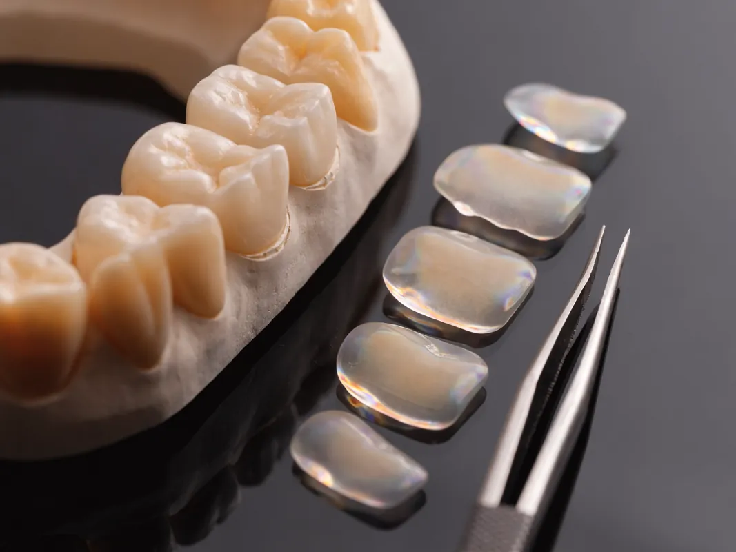
“Natural-looking” is not one measurable outcome. The crown interacts with colour, value, chroma, translucency, fluorescence, surface texture, lustre, incisal effects, shape, contact, symmetry, gingival colour and lighting. Natural neighbouring teeth may have cracks, wear, restorations, dehydration changes or different colours from one another. The patient’s desired shade may conflict with a single-tooth match.
Shade records should include the method, lighting and reference used. Photographs with shade tabs may assist the laboratory, but screens and uncalibrated images distort colour. Whitening natural teeth after making a front crown can create mismatch; if whitening is clinically suitable and desired, sequencing should be discussed before the final shade is selected. Existing restorations do not whiten like natural teeth.
The patient should decide whether the goal is to match current teeth, undertake broader reversible colour treatment first, or accept a planned difference. Adjacent veneers or crowns should not be added casually just to simplify matching. Any irreversible treatment to healthy neighbouring teeth needs its own diagnosis, alternatives and consent.
Abutment and crown materials require balanced discussion
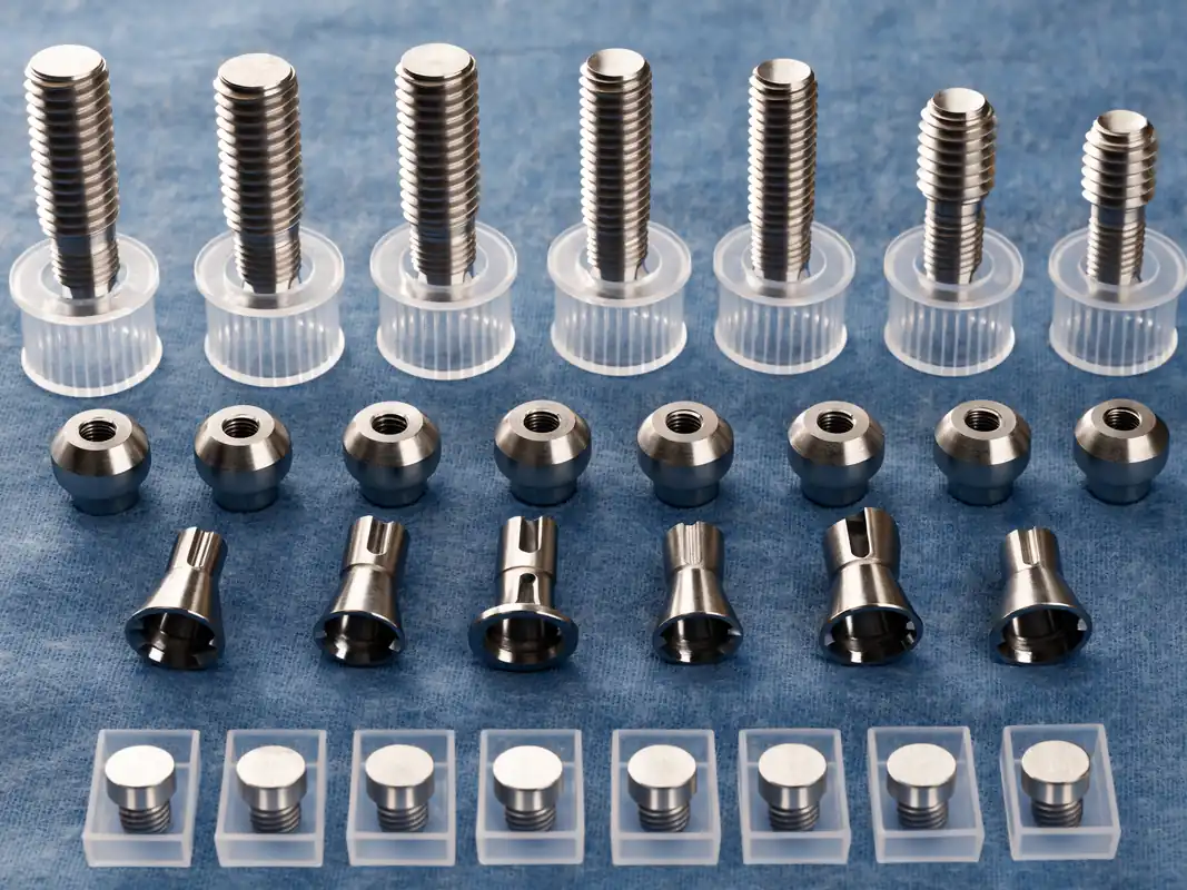
Implant connection, abutment material and crown or framework material affect space, strength, colour, retrievability, repair and component availability. A systematic review of anterior implant abutments reports material-related aesthetic and mechanical findings but does not establish one material as universally superior. Tissue thickness, implant position, connection and restoration design still matter.
Avoid brand adjectives and claims that one ceramic makes a crown invisible. Ask the clinician and laboratory to explain:
- implant system and connection;
- stock or custom abutment and why;
- abutment and crown material;
- screw-retained, cement-retained or other design and its trade-offs;
- how residual cement risk, screw access or colour is managed;
- minimum material requirements and available restorative space;
- repair and replacement pathways;
- whether compatible components are accessible near home.
The choice should be documented without implying a lifetime outcome.
Component, laboratory and shade traceability protect future care
The record should identify what is placed and fitted. Request the implant manufacturer and system, connection, dimensions and site, relevant lot or batch information, healing and restorative components, abutment and screw details, graft or membrane products, crown or framework material, shade references, laboratory prescription and conformity documentation where applicable. Keep operative notes, radiographs, photographs, scans and the final occlusal and tissue records.
“Premium implant” or “European ceramic” is not traceability. Country-of-origin language does not identify a device or prove authenticity. If the planned component changes during treatment, record what was actually used. Future dentists and laboratories may be unable to remove, repair or replace an unidentified restoration safely.
Ask where the laboratory work is made, who checks the prescription, how the clinical team verifies fit and materials, and what happens if the crown needs remanufacture. A laboratory warranty is not a guarantee of biological tissue or aesthetic stability. In Great Britain, relevant MHRA requirements apply to custom-made medical devices; cross-border patients should still obtain intelligible device documentation even where another regulatory system governs manufacture.
Function and occlusion belong in aesthetic consent
Front teeth guide movement, support speech and cut food. An implant lacks the periodontal ligament of a natural tooth, so the clinician must design and review contacts in the context of the whole bite. A central incisor, lateral incisor and canine do not share identical roles. Opposing teeth, deep bite, edge-to-edge relationship, crossbite, wear, parafunction and neighbouring mobility can affect the restoration and provisional plan.
Occlusal adjustment should be documented, not presented as a guarantee against chipping, screw loosening or wear. A night appliance may be considered in selected patients but is not a universal cure and itself needs fit, hygiene and review. Existing jaw pain or temporomandibular symptoms should be assessed separately; replacing a tooth cannot be promised to cure them.
The patient should know which contacts are intended in the provisional and final restoration, what restrictions apply, and who will reassess the bite after settling or if symptoms occur. A crown that photographs well but repeatedly fractures or overloads neighbouring teeth is not a satisfactory plan.
Design the restoration for hygiene and maintenance
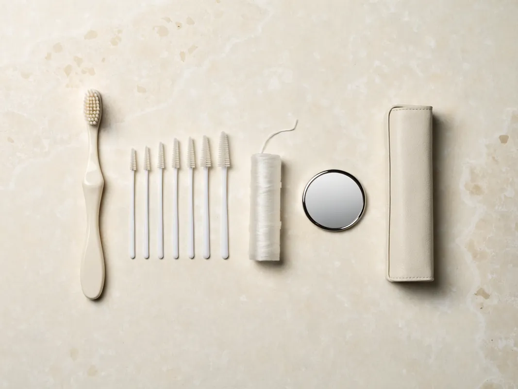
The emergence contour, contact areas and tissue access must allow the patient and dental team to control plaque and examine the site. Over-contoured crowns, deep inaccessible margins or tight tissue architecture can impair cleaning. The patient should be shown the actual brushes, floss or other aids required and demonstrate that they can use them.
Professional maintenance may assess symptoms, plaque, bleeding, probing where appropriate, tissue margin, recession, papilla, occlusion, mobility, screw or component integrity, wear and radiographs when clinically indicated. The EFP peri-implant guideline supports supportive care and prosthetic designs that facilitate hygiene and professional assessment. Maintenance frequency should be individualised rather than promised as one standard interval.
Smoking or nicotine use, active periodontal disease and inconsistent hygiene may increase concern, but communication should be supportive rather than punitive. A plan should address risk modification and realistic care. If the patient cannot clean a proposed contour or cannot access maintenance, another prosthesis may be more appropriate.
Consent must make aesthetic uncertainty visible
GDC Principle 3 requires relevant options, risks, benefits and costs to be explained in a way the patient can understand, with time for questions and recognition that consent is continuing. For a front tooth, the consent discussion should cover both health and appearance. A high surgical survival figure from selected studies is not an individual aesthetic promise.
Material uncertainties may include:
- recession or an uneven gum line;
- incomplete papilla fill or a dark embrasure;
- a visible grey or restorative hue through tissue;
- longer, wider or differently contoured crown proportions;
- imperfect shade, translucency or texture matching;
- pink restorative material or a visible transition;
- tissue changes during healing or continued growth;
- the need for grafting, redesign, repair or another provisional;
- chipping, wear, screw loosening, loss of retention or component problems;
- peri-implant inflammation or disease;
- a plan that must change after extraction or surgery;
- future treatment of neighbouring natural teeth changing the match.
Consent should also compare retention, adhesive bridge, conventional bridge, orthodontic, composite, removable and no-immediate-replacement options. The patient has the right to pause or decline. Travel bookings, deposits and cosmetic expectations must not be used as pressure.
Digital simulations, mock-ups and photographic overlays are communication aids. Label what is a real clinical record, what is a proposed design and what is illustrative. Avoid filters or edited tissue. Obtain separate consent for marketing use of patient images; clinical consent is not advertising consent.
A second opinion is reasonable before irreversible aesthetic-zone treatment
Seek an independent opinion where tooth restorability is disputed, growth may be incomplete, several teeth are proposed for extraction, a major graft is proposed, the gum-risk discussion is vague, the provider promises invisibility or the plan requires treatment of healthy neighbouring teeth merely to match one implant crown.
Provide the second clinician with original radiographs or DICOM files, trauma records, periodontal charting, photographs, orthodontic records, diagnostic set-up and proposed prosthesis. Ask a defined question: Can the tooth be retained? Is growth complete enough for this plan? Are the roots and space suitable? Is the implant position prosthetically driven? Are adhesive or orthodontic alternatives credible? Are tissue and aesthetic risks documented?
Different clinicians may reach different reasonable recommendations. The purpose is to expose assumptions and trade-offs, not to shop for a guarantee. A provider that refuses records or discourages independent review creates a warning sign.
Remote review is provisional, not a final treatment plan
A remote enquiry can collect history, trauma documents, photographs, radiographs, DICOM data, orthodontic records, provisional information and goals. It can identify missing records, urgent symptoms and questions. It cannot palpate tissue, probe periodontal attachment, test pulp or mobility, assess the bite fully, confirm growth, inspect a fracture, verify shade under controlled conditions or predict surgical findings.
Any remote proposal and quote should be labelled provisional. Before sending health data, identify the legal recipient, privacy notice, secure channel, purpose, access and retention arrangements. Compressed messaging images may be inadequate for diagnosis. Obtain and transmit original files through an agreed secure method where required.
At the in-person visit, the clinician should confirm or revise diagnosis, imaging need, tooth prognosis, options, risks and fees. A material change requires explanation and renewed consent. The patient should not accept extra extraction or surgery solely because flights and accommodation are already paid.
Demand an itemised written quote
A front-tooth “implant package” hides the stages that matter. Ask the legal clinical provider for an itemised written quotation that separates:
- consultation and specialist opinions;
- trauma, endodontic, periodontal or orthodontic assessment;
- radiographs, CBCT acquisition and interpretation;
- disease control and preparatory care;
- extraction by site;
- socket management, bone or soft-tissue procedures and materials;
- implant placement and the actual system;
- sedation or anaesthesia where relevant;
- adhesive, removable or other interim tooth;
- implant provisional and adjustments;
- impressions, scans, verification, try-in and shade stages;
- abutment, screw, crown or bridge and laboratory fees;
- records, device documentation and handover;
- planned reviews and maintenance;
- management of foreseeable complications or a changed plan;
- cancellation, postponement, refund and complaint terms;
- travel, accommodation and transfers as separate non-clinical items if purchased.
The quote should state exclusions and which fees cannot be fixed before examination or surgery. Ask what happens if the tooth is retained, immediate placement is abandoned, grafting becomes necessary, loading criteria are not met, a removable provisional is needed, shade remanufacture is requested or another trip is clinically required. Do not assume “remedial treatment” pays for local care, flights, accommodation or time away from work.
Compare like with like. A lower or higher headline price does not establish quality. Compare diagnosis, named clinicians, procedures, products, laboratory, provisional strategy, records, contingency and maintenance.
Cross-border travel must follow the clinical plan
The GDC’s patient guidance on treatment abroad and the NHS treatment-abroad checklist advise checking the provider and qualifications, understanding procedures and costs, planning aftercare, knowing complaint routes and considering insurance and travel implications. Front-tooth treatment may be emotionally urgent, but that does not make a compressed itinerary clinically appropriate.
Before booking, obtain:
- the legal provider name and clinic address;
- each treating clinician’s name, role and registration route;
- the provisional sequence and what could change it;
- a realistic attendance window with contingency for review;
- advice on flying, work, activity and diet from the treating clinician;
- an urgent contact route and local escalation plan;
- arrangements for a companion if sedation or medical needs require one;
- insurance terms and exclusions;
- a willing local dentist or specialist for maintenance and urgent care;
- a plan to transfer full records after each stage.
Hotel and transport arrangements are not clinical evidence and should be kept separate from the healthcare quote. A driver or coordinator cannot decide whether a tooth is restorable, whether a fixture can be loaded or whether the final crown is ready. A return flight must not become a prosthetic deadline.
Local aftercare is part of the original decision
Identify local support before treatment, not after a complication. Ask whether the local practice can examine the planned implant system and restoration, and whether it is willing to provide maintenance without assuming responsibility for the original treatment. Share records promptly. Unknown components or absent operative notes can delay repair.
After each stage, retain:
- updated medical and medicine information;
- diagnosis and procedure notes;
- radiographs and reports;
- implant, graft and component identity;
- provisional and final restoration details;
- laboratory and custom-device documents;
- occlusal and shade records;
- medicines and aftercare instructions;
- review findings and warning signs;
- responsible overseas and local contacts.
Remote photographs can supplement review but cannot replace required clinical examination. A provider should not promise to manage every tissue, bite or component problem through messaging.
Red flags before treatment
Pause and seek independent advice if you encounter:
- extraction recommended without a documented restorability assessment;
- a final plan based only on photographs or one panoramic image;
- an age cut-off used as the only evidence that growth is complete;
- orthodontic root position ignored because the visible gap looks wide enough;
- implant selection before the crown and smile are planned;
- “same-day tooth” used without separating placement, loading and provisional stages;
- a promise that immediate placement preserves the gum line;
- guaranteed papilla fill, invisible margins or a perfect shade match;
- soft-tissue grafting presented as mandatory or risk-free;
- healthy neighbouring teeth proposed for treatment only to make matching easier, without alternatives;
- brand names or “qualified dental technician” language replacing material and laboratory records;
- no adhesive bridge, orthodontic, removable or no-treatment discussion;
- no named clinician, provider, facility or complaint route;
- consent led only by sales staff or an unqualified interpreter;
- refusal to provide DICOM data, reports, component identity or an itemised quote;
- an expiring discount tied to immediate consent;
- no contingency if immediate placement or loading is abandoned;
- no local maintenance or urgent-care plan.
Red flags after trauma, extraction, grafting or implant treatment
Follow the treating clinician’s individual instructions. Difficulty breathing or swallowing, rapidly increasing facial or neck swelling, uncontrolled bleeding, collapse, severe trauma or another emergency requires urgent assessment. Seek prompt professional advice for worsening swelling or pain, fever, pus or foul discharge, persistent vomiting or dehydration, new or persistent altered sensation, wound opening, exposed graft or device, a loose implant or prosthesis, a sudden bite change or inability to clean and eat as instructed.
After trauma, new discolouration, swelling, a sinus tract, increasing mobility, loss of a restoration or another change may require review even if the original injury seemed minor. Follow the trauma team’s schedule and do not substitute an implant sales consultation for monitoring.
The NHS urgent dental guidance explains how to obtain urgent dental help in the UK and identifies situations needing emergency care. Do not wait for an overseas reply when local emergency assessment is required. Notify the original provider when safe and preserve written local findings.
Questions to ask the named clinical team
Use this list at the consultation:
- What is the exact diagnosis and cause of the missing or compromised tooth?
- Can the natural tooth be retained, and has the relevant specialist opinion been obtained?
- What trauma or infection follow-up is still required?
- Is growth complete enough for an implant decision, and how was that assessed?
- Could orthodontics close or redistribute the space?
- Are the adjacent roots positioned safely for the intended restoration?
- What happens if the space is monitored for now?
- Is direct composite repair or reshaping relevant?
- Is a resin-bonded bridge suitable, and what design and maintenance are expected?
- Would a conventional bridge or removable prosthesis be reasonable?
- What is the intended crown position before surgery is planned?
- Can I see the diagnostic set-up and cleanability?
- What does my smile line and lip movement reveal?
- What are the gum phenotype, papilla and ridge risks?
- Which imaging is justified, and who interprets it?
- Why is immediate, early or delayed placement proposed?
- What would make that placement plan change during treatment?
- Is bone or soft-tissue augmentation proposed, and why?
- What materials and donor sources may be used?
- What criteria must be met before loading?
- What temporary tooth will I have if the implant cannot be loaded?
- How are provisional and final restorations separated?
- What shade, gum-line and papilla differences cannot be ruled out?
- What implant, abutment and crown materials are proposed?
- How will component, laboratory and shade details be recorded?
- How is the restoration designed for function and cleaning?
- What is included and excluded in the itemised quote?
- Which costs follow if the preferred sequence changes?
- Who provides urgent care and maintenance near my home?
- Can I take the full records for an independent second opinion?
A practical patient decision checklist
Before consent, confirm that you can answer yes to the following:
- I know whether the tooth can reasonably be retained.
- Trauma, infection and periodontal disease have a documented plan.
- Growth has been considered where relevant.
- Space, roots, smile, gum, bone, function and hygiene were assessed.
- I have compared implant, adhesive bridge, conventional bridge, orthodontic, composite, removable and no-immediate-replacement options.
- I understand why the proposed placement timing was selected.
- I understand that placement, loading, provisional and final stages are different.
- I know the fallback if immediate placement or loading is not possible.
- I understand papilla, gum-line, shade and contour uncertainty.
- I know the proposed material and component record.
- I have an itemised quote and change-of-plan terms.
- I know the named provider and clinicians.
- I have local aftercare and urgent-care routes.
- I have time to seek a second opinion and can postpone without clinical pressure.
Evidence and further reading
These sources inform the framework. They do not diagnose an individual or provide a personal probability of aesthetic success.
- American Association of Endodontists, AAE Position Statement – Implants, accessed 29 August 2026: preserve restorable natural teeth, compare retention and replacement, and discuss complications and consent. https://www.aae.org/specialty/wp-content/uploads/sites/2/2019/04/Implants_PositionStatement_v2.pdf
- International Association of Dental Traumatology guideline for fractures and luxations of permanent teeth, published 2020 and accessed 29 August 2026: trauma diagnosis, imaging, documentation, pulp assessment, instructions and follow-up. https://www.aapd.org/globalassets/media/policies_guidelines/e_fractures.pdf
- American Academy of Pediatric Dentistry, Best Practices: Periodontal Conditions, current document accessed 29 August 2026: conservative missing-tooth care during growth and multidisciplinary implant assessment. https://www.aapd.org/globalassets/media/policies_guidelines/bp_periotherapy25.pdf
- Systematic review of implant and ankylosed-tooth infraposition related to craniofacial growth, accessed 29 August 2026: long-term positional uncertainty in the anterior maxilla. https://pubmed.ncbi.nlm.nih.gov/32835562/
- British Orthodontic Society, My Missing Teeth, accessed 29 August 2026: orthodontic space closure or creation and restorative alternatives. https://bos.org.uk/patients/my-missing-teeth/
- American Dental Association and American Academy of Oral and Maxillofacial Radiology clinical recommendations, published 2026 and accessed 29 August 2026: examination-led imaging selection and CBCT use in implant planning. https://pubmed.ncbi.nlm.nih.gov/41500761/
- ITI consensus, Clinical Performance of Immediately Placed and Immediately Loaded Single Implants in the Esthetic Zone, accessed 29 August 2026: selected clinical viability and recognised complications. https://academy.iti.org/academy/consensus-database/consensus-statement/-/consensus/clinical-performance-of-immediately-placed-and-immediately-loaded-single-implants-in-the-esthetic-zone/2304
- Supporting systematic review of immediate placement and loading in the aesthetic zone, published 2023 and accessed 29 August 2026: evidence boundaries and selected study populations. https://pubmed.ncbi.nlm.nih.gov/37750531/
- Systematic review of papilla height around single-tooth implants, accessed 29 August 2026: adjacent-tooth attachment and contact-to-bone relationships with limited evidence. https://pubmed.ncbi.nlm.nih.gov/29498124/
- Osteology Foundation consensus report on peri-implant soft-tissue aesthetics, accessed 29 August 2026: papilla uncertainty for single and adjacent implants. https://pubmed.ncbi.nlm.nih.gov/29498131/
- Systematic review of connective-tissue grafts around immediately placed and restored aesthetic-zone implants, accessed 29 August 2026: case-specific soft-tissue evidence. https://pubmed.ncbi.nlm.nih.gov/32311199/
- Systematic review of anterior single-retainer resin-bonded prostheses, published 2026 and accessed 29 August 2026: conservative bridge evidence and limitations. https://pubmed.ncbi.nlm.nih.gov/42457533/
- Systematic review of anterior implant abutment materials, accessed 29 August 2026: aesthetic, mechanical and biological trade-offs without a universal material winner. https://pubmed.ncbi.nlm.nih.gov/34494174/
- European Federation of Periodontology S3 guideline on peri-implant diseases, published 2023 and accessed 29 August 2026: disease prevention, cleanable prostheses and supportive care. https://pubmed.ncbi.nlm.nih.gov/37271498/
- General Dental Council Principle 3, Obtain valid consent, accessed 29 August 2026: options, risks, benefits, costs, time and continuing consent. https://standards.gdc-uk.org/pages/principle3/principle3
- General Dental Council Principle 4, Maintain and protect patients’ information, accessed 29 August 2026: complete clinical, radiographic, photographic and laboratory records. https://standards.gdc-uk.org/pages/principle4/principle4
- General Dental Council Principle 7, Work with colleagues in patients’ interests, accessed 29 August 2026: competence, collaboration and referral. https://standards.gdc-uk.org/pages/principle7/principle7
- Medicines and Healthcare products Regulatory Agency, custom-made medical devices in Great Britain, updated 20 July 2026 and accessed 29 August 2026: documentation and responsibilities for relevant devices. https://www.gov.uk/government/publications/custom-made-medical-devices/custom-made-devices-in-great-britain
- General Dental Council, Going abroad for dental treatment, accessed 29 August 2026: provider verification, records, costs, aftercare and complaints. https://www.gdc-uk.org/standards-guidance/information-for-patients-public/going-abroad-for-dental-treatment
- NHS, Treatment abroad checklist, accessed 29 August 2026: travel, insurance, follow-up and contingency questions. https://www.nhs.uk/using-the-nhs/healthcare-abroad/going-abroad-for-treatment/treatment-abroad-checklist/
- NHS, How to find an NHS dentist in an emergency, accessed 29 August 2026: urgent dental access and emergency warning signs. https://www.nhs.uk/nhs-services/dentists/how-to-find-an-nhs-dentist-in-an-emergency/
The evidence supports a diagnosis-led, tooth-preserving and prosthesis-led decision with explicit aesthetic uncertainty. It does not support promises of an invisible crown, guaranteed papilla, fixed same-day loading or a universal front-tooth implant protocol.