A broken front tooth can mean a small enamel chip, an exposed pulp, a fracture extending below the gum, a loosened or displaced tooth, a root injury, or a tooth that has been completely knocked out. Those injuries do not share one repair. A photograph can help describe what happened, but it cannot determine the depth, pulp status, root condition, supporting-tissue injury or restorability.
Urgent triage comes before cosmetic planning. If the injury is recent, the first priority is appropriate local dental assessment—not a flight, remote quotation or choice between a veneer and an implant. Once urgent needs are managed, a responsible plan can compare tooth-preserving repairs, follow-up, definitive restoration and replacement only where the tooth cannot reasonably be maintained.
This guide is not an emergency service and does not diagnose a particular tooth. It provides a structured way to ask questions, preserve evidence, compare options and avoid turning “broken front tooth repair Turkey” into a standard cosmetic package. It publishes no fixed repair, time, price, outcome or colour-match promise. Those require individual clinical assessment by the responsible clinician.
First actions after a chip or break
If the tooth has just broken:
- contact a local dentist or appropriate urgent dental service;
- save a detached fragment if it can be found;
- handle the fragment carefully and keep it from drying out;
- follow the local dental service's instructions about storage and transport;
- report bleeding, pain, sensitivity, tooth movement, displacement, altered bite, facial injury, loss of consciousness, vomiting, double vision or other symptoms;
- avoid testing the tooth repeatedly or using it to bite;
- use only medicine that is safe for the person and consistent with professional advice and the label;
- do not postpone urgent assessment to obtain an overseas cosmetic estimate.
The [NHS chipped, broken or cracked tooth page](https://www.nhs.uk/conditions/chipped-broken-or-cracked-tooth/) advises taking a broken fragment to the dentist and notes that milk or saliva can be used for storage. The fragment may sometimes be reattached. The same page directs people to dental rather than general medical treatment for the tooth itself.
For UK users, the [NHS urgent and emergency dental page](https://www.nhs.uk/nhs-services/dentists/how-to-find-an-nhs-dentist-in-an-emergency/) distinguishes urgent dental needs from conditions requiring emergency medical care. Serious facial or jaw injury, uncontrolled heavy mouth bleeding, severe swelling affecting breathing, or head injury symptoms need the emergency route described there. People elsewhere should use the equivalent local service.
These steps are general triage, not a personal protocol. A clinician who can assess the injury should determine what happens next.
Describe the event before naming the restoration
The history affects diagnosis and follow-up. Tell the clinician:
- when and how the injury occurred;
- whether the tooth or fragment was contaminated, dry or stored;
- whether the tooth moved, changed position or affected the bite;
- whether there was loss of consciousness or another head or facial injury;
- whether the lip, tongue or cheek was cut and whether a fragment may be embedded;
- current pain, temperature sensitivity, tenderness, swelling, bleeding, numbness or colour change;
- previous trauma, root canal treatment, restorations, orthodontics or gum treatment involving the tooth;
- medical conditions, medicines, allergies and tetanus-related questions where a treating professional considers them relevant;
- whether the tooth is permanent or primary and whether growth is complete.
Do not assume that a visually small chip is the only injury. Supporting tissues can be injured at the same time. Conversely, a dramatic-looking missing corner may leave a maintainable tooth with conservative options. Diagnosis has to connect the story with clinical findings.
Injury classification changes the pathway
Terms matter because a crown fracture, crown-root fracture, root fracture, luxation and avulsion describe different problems.
| Injury category | What it broadly means | Why the distinction matters |
|---|---|---|
| Enamel chip | Limited loss of hard outer tissue | May need smoothing, bonding, fragment use or monitoring rather than broad coverage |
| Enamel-dentine fracture | More tooth structure lost without visible pulp exposure | Dentine protection, restoration and pulp follow-up may be relevant |
| Complicated crown fracture | Fracture with pulp exposure | Pulp status, root maturity, timing and vital-pulp or endodontic decisions need clinician assessment |
| Crown-root fracture | Fracture extends below the gum toward the root | Access, depth, periodontal relationship and restorability become central |
| Root fracture | Injury affects the root | Position, mobility, pulp and supporting tissues need trauma-specific management and review |
| Luxation | Tooth is loosened or displaced without complete avulsion | Repositioning, stabilisation and supporting-tissue follow-up may be time-sensitive |
| Avulsion | Permanent tooth is completely knocked out | Immediate emergency management differs fundamentally from restoring a chipped crown |
| Longitudinal crack | Crack may progress along crown or root and may not be caused by one acute event | Extent and restorability can be difficult to diagnose; treatment depends on the actual crack |
The [International Association of Dental Traumatology guideline for fractures and luxations of permanent teeth](https://www.aapd.org/research/oral-health-policies--recommendations/guidelines-for-the-management-of-traumatic-dental-injuries-1-fracture-and-luxations-or-permanent-teeth/) is an evidence-and-consensus resource for clinicians. It emphasises diagnosis, treatment planning and follow-up and explicitly does not guarantee a favourable outcome. Its acute-trauma pathways cannot be compressed into a veneer-versus-crown sales table.
Diagnosis needs history, examination and appropriate imaging
A responsible assessment may include inspection, palpation, mobility and displacement checks, bite assessment, periodontal findings, pulp sensibility tests, photographs and radiographs selected for the clinical question. The required set depends on the injury, age, symptoms and earlier records. More imaging is not automatically better, and one panoramic image cannot answer every anterior-tooth question.
Ask the clinician to document:
- the injury classification;
- tooth and surface involved;
- remaining tooth structure and fracture margins;
- whether the pulp is exposed or otherwise at risk;
- mobility, displacement and supporting-tissue findings;
- root development and relevant root findings;
- periodontal relationship of any subgingival fracture;
- adjacent and opposing tooth findings;
- restorability and information still required;
- baseline tests and the planned follow-up tests;
- which images were reviewed and who interpreted them.
If a crack is suspected rather than a simple traumatic chip, diagnosis can be challenging. The [European Society of Endodontology position statement on longitudinal cracks and fractures](https://pubmed.ncbi.nlm.nih.gov/39840523/) addresses aetiology, presentation and management based on evidence and expert consensus. It does not create one online rule for every crack.
Pulp status can change over time
The pulp is living tissue inside the tooth. Trauma may expose it directly, disrupt its blood supply or cause changes that appear later. A single early sensibility result does not always establish the long-term status, particularly after trauma. Follow-up is diagnostic, not a ceremonial check.
The written plan should distinguish:
- current pulp-related findings;
- the reliability and limits of tests at that stage;
- whether the root is mature or still developing;
- symptoms or signs that require earlier review;
- what would trigger vital-pulp treatment, root canal treatment or another decision;
- how an interim restoration permits monitoring and access;
- who owns follow-up after the person travels.
Root canal treatment is a separate diagnosis and should not be added automatically because a front tooth broke. Conversely, an apparently successful cosmetic repair does not remove the need to monitor the tooth where the injury creates pulp or supporting-tissue risk.
Establish restorability before extraction
Before comparing veneer, crown or implant, ask whether the tooth or fragment can be maintained and what evidence supports that conclusion. Restorability includes more than “a root is visible on the scan”. It may involve the fracture depth, remaining sound tissue, ferrule possibilities where relevant, pulp and root status, periodontal support, biological relationships, bite, moisture control, access for restoration, aesthetics, maintenance and the consequences of proposed procedures.
Ask for a restorability statement:
| Question | Evidence to request |
|---|---|
| What structure remains? | Tooth map, fracture description, photographs and clinical findings |
| Is the fracture accessible? | Margin location and periodontal relationship |
| What is the pulp/root status? | Tests, images, diagnosis and uncertainty |
| Can the fragment be used? | Condition, fit, storage history and restorative assessment |
| What conservative options exist? | Smoothing, reattachment, direct repair or monitoring where appropriate |
| What additional procedures are proposed? | Purpose, alternatives, risks and effect on prognosis |
| What would make the tooth non-restorable? | Patient-specific finding, not a generic statement |
| Is extraction reversible? | No; request an independent opinion where doubt remains |
There is no universal repair hierarchy that moves automatically from veneer to crown to implant. The least invasive suitable option may preserve future choices, but “least invasive” must still address the actual injury and support a maintainable result.
Fragment reattachment can be an option
When a usable fragment exists, fragment reattachment may preserve the tooth's original shape, texture and optical features. It is not always feasible and it is not failure-proof. Suitability depends on the fracture, fragment condition, pulp and root issues, ability to isolate and bond, bite, age and the clinician's judgement.
The [systematic review of fragment reattachment after complicated crown-root fractures](https://pubmed.ncbi.nlm.nih.gov/32813931/) found mainly case reports and case series. It described reattachment as a possible option under favourable clinical conditions but also reveals the evidence limitations. A case series cannot provide a personal lifespan promise.
Ask:
- whether the fragment is complete and fits;
- whether dehydration, contamination or damage affects its use;
- what pulp or root treatment is separately indicated;
- how the fracture line is accessed and isolated;
- whether additional preparation is proposed and why;
- what bite protection and follow-up are needed;
- what repair is possible if the fragment debonds or fractures;
- which alternative preserves the most tissue if reattachment is unsuitable.
Keep the fragment even if reattachment seems unlikely. The treating dentist should decide after examining it.
Direct composite is a distinct conservative repair
Direct composite can rebuild missing front-tooth structure without manufacturing an indirect restoration. Its suitability depends on the size and position of the defect, enamel and dentine available for bonding, isolation, pulp status, bite, colour, operator skill and maintenance expectations.
The plan should explain whether composite is intended as an emergency seal, provisional repair or longer-term restoration. Those are different roles. Ask how the contour, contact, edge position, polish and bite will be checked, and how future repair or replacement would work.
Composite can stain, wear, chip or need repolishing or repair; those possibilities do not make it unsuitable. They belong in the comparison with the additional tooth preparation, laboratory stages and repair patterns of indirect options.
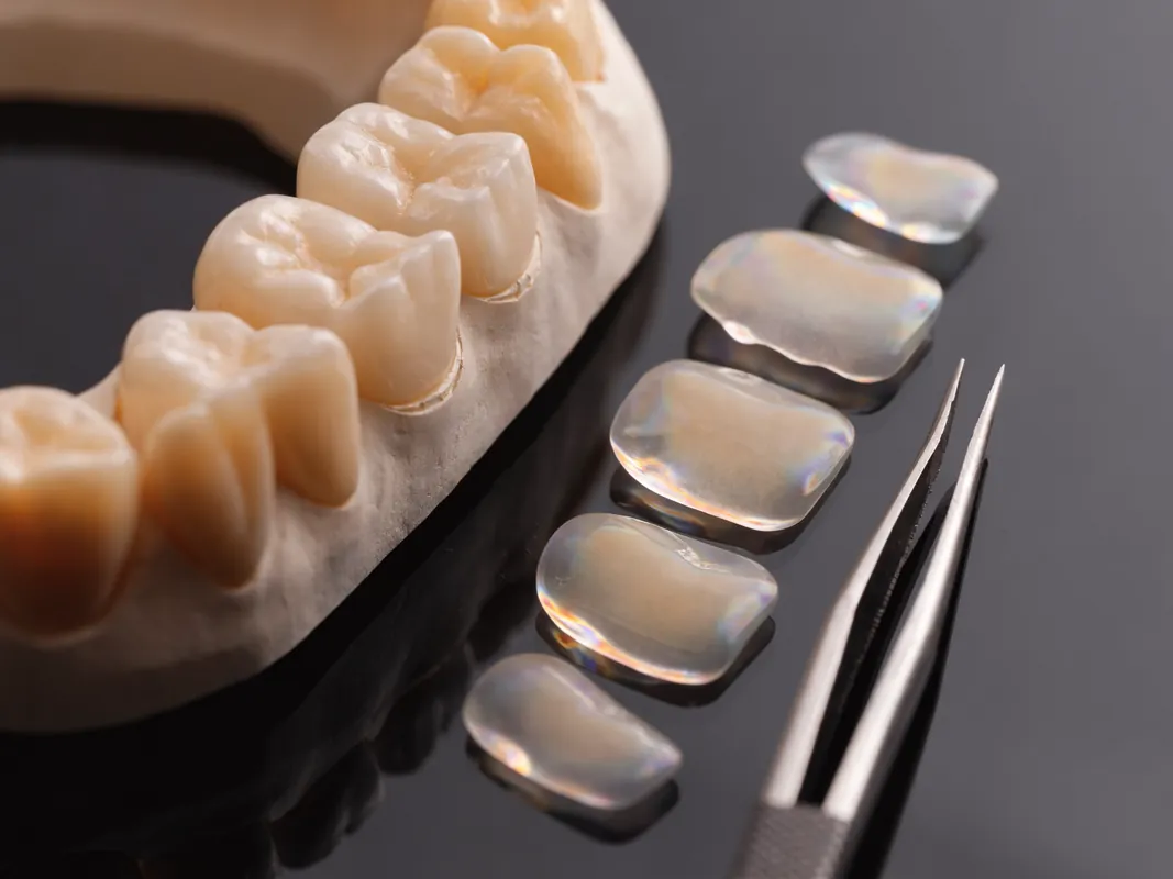
A veneer is not the default repair
A veneer may be discussed for selected defects or later aesthetic management, but veneer is not the default repair for a broken front tooth. The clinician must assess whether the defect, enamel available for bonding, pulp and root condition, margin position, bite and colour goals support it. A veneer does not stabilise every crack, treat infection or replace a missing root.
Ask:
- which surface and structure the veneer would cover;
- how much preparation is proposed and whether a no- or minimal-preparation alternative is realistic;
- why fragment reattachment or direct composite is unsuitable;
- whether the tooth needs trauma follow-up before an irreversible elective veneer;
- how the margin relates to the fracture and gum tissues;
- how shade changes in the injured tooth would be managed;
- what happens if later endodontic access or repair is needed.
The related dental veneer guide covers broader veneer planning. This page owns the trauma-to-repair decision.
A crown is not the default repair
A crown covers much more tooth structure than a localised direct repair. It may be considered where the remaining tooth, restoration, function and prognosis support full coverage, but crown is not the default repair simply because the missing piece is visible.
Ask the restorative clinician to document:
- the reason full coverage is proposed;
- how much maintainable tissue remains;
- whether the margin can be placed and maintained appropriately;
- whether a core or post is proposed and its separate indication;
- which more conservative options were assessed;
- how the restoration preserves future endodontic access where relevant;
- material and laboratory prescription;
- bite, contact, shade and maintenance plan.
Root canal treatment does not automatically make every tooth need the same crown, and a crown does not treat an undiagnosed pulp or root problem. The root canal and crown sequencing guide separates those decisions.
Crown-root fractures may need a multidisciplinary decision
When the fracture extends below the gum, the team may need to consider periodontal access, restorative isolation, pulp/root treatment and the final margin together. Options discussed in individual cases can include fragment use, direct or indirect restoration, orthodontic movement, surgical exposure, extraction or another strategy. Listing an option does not mean it is suitable.
Request one shared map showing:
- the deepest fracture extent that can be established;
- supporting-tissue and root findings;
- whether proposed exposure or movement changes crown-root proportions or aesthetics;
- who owns periodontal, endodontic, orthodontic and restorative stages;
- which stage remains provisional;
- how each option affects time, cost, future maintenance and travel;
- the no-treatment option and consequences of delay.
A package timetable should not decide whether the tooth is restorable.
Extraction and replacement are separate decisions
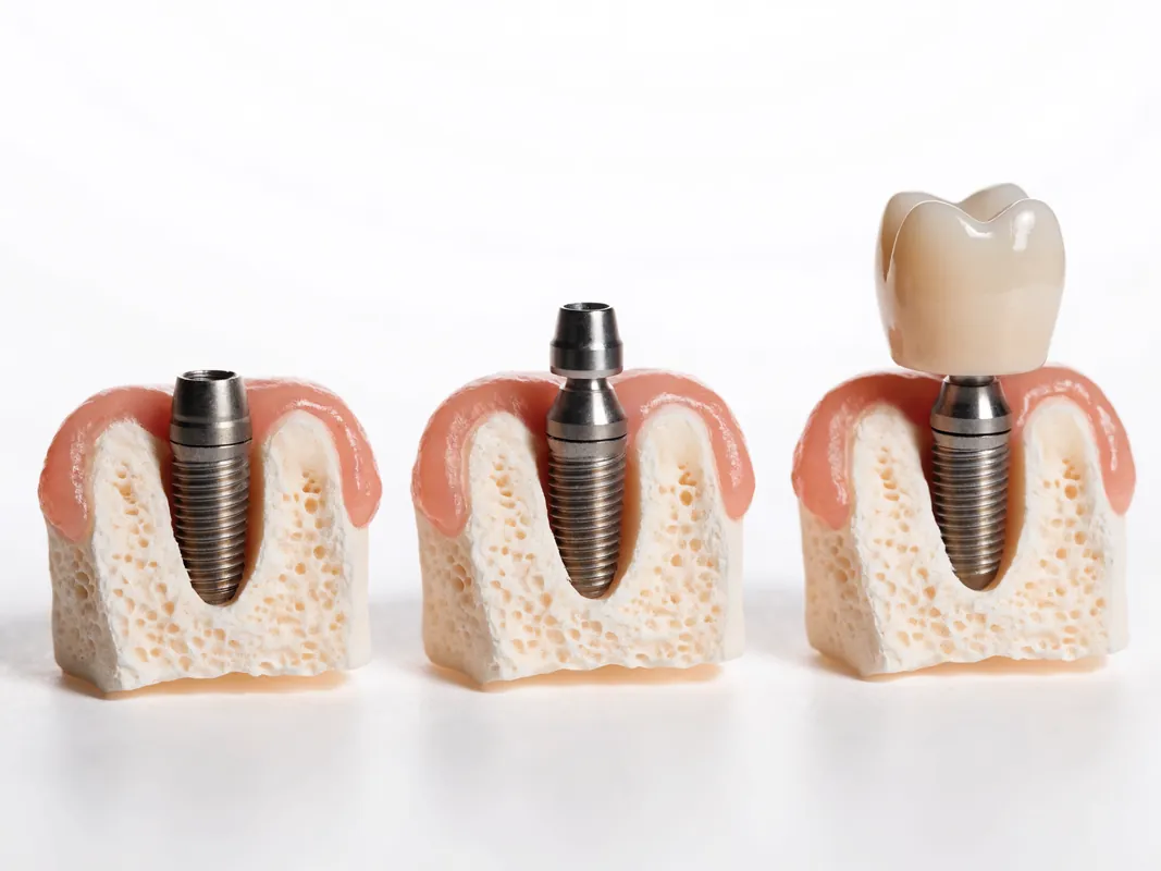
If the tooth is judged non-restorable, ask for the finding and an independent opinion before extraction when practical. Extraction is one decision; the replacement method and timing are separate decisions. Implant is not an emergency substitute for diagnosis or a guaranteed way to reproduce a natural front tooth.
Replacement alternatives may include no immediate replacement, a removable temporary option, an adhesive or conventional bridge, orthodontic space management, an implant-supported crown, or another case-specific plan. They involve trade-offs in surgery, neighbouring teeth, growth, bone and soft tissue, timing, appearance, cleaning, maintenance and repair.
For an implant proposal, ask:
- whether growth and age have been considered;
- what preserves appearance and function during healing;
- what bone and soft-tissue findings support the site plan;
- whether immediate placement or loading is proposed and on which conditions;
- what happens if those conditions are not met;
- how tooth position, gum level and papillae are assessed;
- which system and components are used and recorded;
- how local aftercare and future component access work.
Do not accept “broken below the gum” as a complete extraction diagnosis. The actual fracture, root, periodontal and restorative findings matter.
Growth and age change the comparison
Traumatic front-tooth injuries are common in children and young people, but adult cosmetic pathways cannot simply be copied to a growing patient. Root maturity can affect pulp-management decisions. Ongoing facial and dental development can affect definitive restorative and implant timing. Primary and permanent teeth also have different trauma pathways.
The responsible dental team should identify the tooth type, stage of root development and growth considerations and involve an appropriately experienced clinician where needed. A temporary or repairable solution may deliberately preserve future options. That is not incomplete care; it can be a planned stage.
Parents and carers should receive clear information, but the young person's involvement, understanding and consent or assent need to follow applicable professional and legal requirements. A travel offer should never compress those safeguards.
Aesthetic matching is a process, not a promise
Front-tooth restoration involves shade, translucency, texture and shape as well as position, edge character, surface reflection, gum relationship and how the tooth appears in different light. The adjacent tooth may also have been injured. The damaged tooth can change colour over time, and restorative materials behave differently.
Ask how the team records:
- photographs with useful colour references;
- hydration state, because a dry tooth can look different;
- shade and character of neighbouring teeth;
- edge position and symmetry without assuming identical teeth;
- surface texture and gloss;
- provisional feedback;
- laboratory prescription and authorisation of changes;
- limitations caused by the remaining tooth, substrate, material or tissue.
A technician and clinician can aim for a harmonious repair, but no responsible plan can promise that it will be visually undetectable in every light and angle. Consent should separate the clinical repair from optional treatment of otherwise healthy neighbouring teeth.
Provisional repair has a defined role
A provisional repair may protect exposed tissue, restore a safe contour, support appearance or function, and preserve options while diagnosis and follow-up continue. It should have a documented purpose, limitations, review route and contingency.
Ask whether the provisional:
- is intended for hours, a diagnostic stage or a longer interim period without assuming a fixed schedule;
- permits pulp and trauma monitoring;
- changes eating or bite instructions;
- can be repaired locally;
- must be removed for a later stage;
- is included in the quotation;
- affects the definitive design;
- has warning signs requiring earlier assessment.
“Temporary” does not mean clinically unimportant. A poor contour, unstable restoration or lost seal may need prompt review.
Follow-up is diagnostic
Trauma follow-up can assess symptoms, pulp and supporting-tissue changes, root development, mobility, colour, restoration integrity, gum health and radiographic changes where justified. The schedule and tests should follow the injury and clinician's assessment, not a generic package interval.
Before travelling, ask:
- which reviews are time-sensitive;
- who will perform them;
- which baseline records are needed for comparison;
- how results are shared between providers;
- what findings trigger a new diagnosis or treatment;
- whether a local dentist has accepted the proposed role;
- what happens if the restoration fails or symptoms develop abroad.
A tooth can look acceptable while a biological problem develops, and a restoration can need repair while the tooth remains maintainable. Separate those outcomes.
Travel should not delay urgent care
Do not wait for a trip to have acute trauma assessed. Early local diagnosis may preserve treatment options, manage pain or infection, stabilise an injury and create baseline records. Later travel for elective repair is a separate decision.
If overseas treatment is considered after stabilisation, the plan should state:
- what urgent care is already complete;
- what remains diagnostic or provisional;
- whether travel timing conflicts with follow-up;
- who owns the tooth before, during and after the trip;
- what local clinician will do if symptoms change;
- how flights, accommodation and payments change if the clinical plan changes;
- whether travel insurance addresses planned dental care and complications.
Never substitute an airport date for a clinical decision gate.
Compare options with a decision matrix
The matrix below is a prompt, not a recommendation:
| Option | Questions to answer before choosing |
|---|---|
| Smoothing or monitoring | Is the defect limited, safe and free from deeper injury requiring treatment? |
| Fragment reattachment | Is the fragment usable, can the field be isolated, and what follow-up is required? |
| Direct composite | Is suitable structure available for bonding, and how repairable is the design? |
| Veneer | Why is broader indirect coverage justified, and what enamel and trauma monitoring remain? |
| Crown | Why is full coverage appropriate, how much tissue remains, and are pulp/root decisions separate? |
| Multidisciplinary salvage | Can subgingival fracture management create a maintainable restorative situation, and at what cost or risk? |
| Extraction | Which finding makes the tooth non-restorable, and has an independent opinion been considered? |
| Bridge | What happens to neighbouring teeth, cleaning, repair and future options? |
| Implant crown | Are site, growth, tissue, timing, loading and aftercare conditions supported? |
| No treatment or delay | What risks, symptoms and future limitations require monitoring? |
There is no universal repair hierarchy. Compare preservation, diagnosis, reversibility, evidence, maintenance and future options before appearance or trip length.
Make the quotation itemised
An itemised quotation should identify:
- assessment and diagnostic records;
- urgent stabilisation already completed or still required;
- fragment reattachment or direct repair where proposed;
- pulp, root canal or other endodontic care only where separately diagnosed;
- periodontal, orthodontic or surgical stages where proposed;
- provisional restoration and repairs;
- definitive restoration, material and laboratory;
- extraction and temporary replacement where applicable;
- implant, bridge or removable replacement as separate choices;
- review appointments and diagnostic follow-up;
- records and handover;
- local aftercare and urgent assessment boundaries;
- travel services under a separate contract;
- exclusions, conditional items, payment stages and cancellation;
- changed-plan consent and price approval.
If the in-person diagnosis changes the repair, require an updated dated plan before proceeding. A deposit should not become consent for a more destructive option.
Records and handover prevent guesswork
Request copies of the history, injury classification, examination, tests, photographs, radiographs and interpretations, urgent treatment, materials, pulp or root diagnosis, restoration and laboratory prescription, medicine record, consent discussions, review findings and unresolved concerns.
The [GDC consent standard](https://standards.gdc-uk.org/pages/principle3/principle3) emphasises explaining relevant options and costs, checking understanding and renewing consent when treatment or price changes. The [GDC record standard](https://standards.gdc-uk.org/pages/principle4/principle4) lists radiographs, consent forms, photographs, models, laboratory prescriptions and referrals among patient records where available. These are UK professional comparison frameworks, not statements of Turkish law.
Ask which legal and professional rules apply to the provider treating the person. Records should be supplied in a usable form and transferred securely with consent. A local clinician should not have to reconstruct the injury history from social-media messages.
Red flags in a broken-tooth proposal
Pause when:
- a final repair is selected from a photograph alone;
- urgent local assessment is discouraged so a package can be sold;
- the tooth is called hopeless without a restorability statement;
- a veneer or crown is presented as mandatory for every chip;
- root canal treatment is added without a separate diagnosis;
- extraction and implant placement are treated as one automatic step;
- a front implant is promised to match perfectly;
- the plan ignores growth, root maturity or trauma follow-up;
- the provider cannot name the responsible clinician;
- the quotation hides provisional, endodontic, periodontal or laboratory items;
- records and material information will not be supplied;
- aftercare is limited to sending another photograph;
- travel dates determine a clinical decision.
A red flag is a prompt to obtain evidence. If a critical question remains unanswered, obtain urgent local care, an independent opinion or another plan.
Questions to send a provider
- What injury classification and restorability finding supports the proposed repair?
- Which examination, tests and images have been reviewed, and what remains unknown?
- Is there evidence of luxation, root injury or supporting-tissue trauma as well as crown damage?
- Can the fragment be assessed for reattachment?
- Is direct composite a suitable conservative option?
- Why is a veneer or crown proposed instead of a smaller repair?
- Is pulp treatment indicated now, or is monitoring required?
- What finding would trigger root canal treatment later?
- If the fracture is subgingival, which salvage options were considered?
- Which finding makes extraction necessary, if proposed?
- Which replacement options and no-treatment option were discussed?
- How do growth and age affect definitive timing?
- What is the provisional repair's role and limitation?
- How will shade, translucency, texture and shape be recorded and reviewed?
- Who owns trauma follow-up after travel?
- Which records and material details will be supplied?
- How is the quotation divided by diagnostic, provisional and definitive stage?
- What happens to consent, cost and travel if the diagnosis changes?
Sources and evidence limits
These sources were checked on 29 August 2026. They support triage, classification, decision questions and evidence boundaries. They do not diagnose an individual tooth, endorse a provider or guarantee a repair.
- [NHS: chipped, broken or cracked tooth](https://www.nhs.uk/conditions/chipped-broken-or-cracked-tooth/) — public guidance to see a dentist, preserve a detached fragment and understand broad treatment categories.
- [NHS: urgent and emergency dental access](https://www.nhs.uk/nhs-services/dentists/how-to-find-an-nhs-dentist-in-an-emergency/) — UK triage boundaries for dental injury and serious head, face, bleeding, swelling or breathing concerns.
- [IADT fractures and luxations guideline, hosted by AAPD](https://www.aapd.org/research/oral-health-policies--recommendations/guidelines-for-the-management-of-traumatic-dental-injuries-1-fracture-and-luxations-or-permanent-teeth/) — professional evidence-and-consensus pathways for permanent-tooth trauma, with explicit outcome limitations.
- [European Society of Endodontology position statement on cracks and fractures](https://pubmed.ncbi.nlm.nih.gov/39840523/) — evidence and expert consensus for longitudinal crack presentation and management.
- [Systematic review: fragment reattachment after complicated crown-root fracture](https://pubmed.ncbi.nlm.nih.gov/32813931/) — evidence that reattachment may be considered in favourable conditions, with an evidence base dominated by reports and series.
- [GDC Principle 3: obtain valid consent](https://standards.gdc-uk.org/pages/principle3/principle3) — UK comparison framework for options, costs, understanding and changed-plan consent.
- [GDC Principle 4: maintain and protect patient information](https://standards.gdc-uk.org/pages/principle4/principle4) — UK comparison framework for accurate, accessible and secure records.
The IADT guideline is designed to assist clinicians and patients, not to replace professional judgement. Evidence strength differs across injury types and repairs. A personal decision still requires current findings, responsible clinicians, valid consent and follow-up.
Final decision rule
Treat the injury before shopping for a cosmetic category. Preserve a fragment, obtain appropriate local assessment, classify the injury, document pulp/root/supporting-tissue findings, test restorability, compare conservative repairs, and separate extraction from replacement. If elective travel remains useful after stabilisation, carry the records and keep clinical timing independent of flights.
The best documented plan is not necessarily the most extensive one. It is the plan that explains why the proposed repair fits the actual injury, what remains uncertain, how the tooth will be monitored and what happens if the diagnosis changes.