Tetracycline staining is one possible cause of intrinsic tooth discolouration. It is not a diagnosis that can be confirmed from a filtered smile photograph, and it does not make veneers inevitable. Surface stain, caries, enamel developmental conditions, fluorosis, trauma, pulp disease, ageing, restorative materials and several other causes can produce yellow, brown, grey, banded or uneven teeth. The first task is to identify what changed, which tissues are involved and whether oral disease needs treatment. The second is to find the least destructive reasonable way to meet the patient’s goal.
The old claim that tetracycline-stained teeth “cannot be whitened” is too absolute. Clinical trials report measurable colour change in some tetracycline-stained teeth with professionally supervised extended whitening protocols, while response, burden, sensitivity, stability and acceptability vary. That does not make whitening suitable for everyone or promise complete correction. It means whitening belongs in a real option discussion before sound tooth tissue is removed.
Veneers and crowns also do not guarantee masking. The underlying tooth colour, preparation, remaining enamel and dentine, restorative material, thickness, opacity, cement, laboratory process, lighting, shade target and surrounding teeth interact. More opacity can hide a dark substrate but may reduce depth and translucency. More material space may require more tooth reduction. A “bright” result can still look flat, opaque or mismatched. These are clinician-and-laboratory decisions with trade-offs, not a stock recipe.
This guide is evidence-led information for adults researching tetracycline-stained teeth, intrinsic discolouration, whitening, composite or veneers, including people considering care abroad. It is not a personal diagnosis, prescription, quotation, timetable, material selection or result promise. The named dentist must examine the mouth, diagnose the cause, establish health and suitability, explain alternatives, obtain continuing consent and remain responsible for care. The laboratory works to a clinical prescription; it does not diagnose the patient.
Start with the cause, not the label “tetracycline teeth”
Ask who made the diagnosis and on what evidence. Was there a reliable history of a tetracycline-class medicine during tooth development? Which teeth are affected? Does the distribution correspond with the development of those teeth? Is the colour generalised or limited to one tooth? Are there horizontal bands, diffuse changes, surface deposits, white or brown patches, cracks, restorations, decay or a non-vital tooth? Has the appearance been stable since eruption, or did it change recently?
A patient may not know the exact medicine, dose, age or duration, and family memory can be wrong. Historical exposure can support the diagnosis but does not replace examination. Conversely, not remembering exposure does not prove another cause. The dentist should document the degree of confidence and any differential diagnoses rather than use a severity label as an automatic veneer prescription.
A useful problem list separates:
- extrinsic surface stain from intrinsic colour;
- developmental discolouration from colour acquired after eruption;
- enamel, dentine, pulp and restorative causes;
- one-tooth changes from generalised patterns;
- colour concerns from shape, alignment, wear and gum concerns;
- healthy tissue from caries, cracks, erosion, inflammation or other disease;
- the patient’s desired change from what treatment can reasonably alter;
- reversible options from additive and subtractive restorations;
- known findings from uncertainty that requires testing or monitoring.
“Grade” systems can help clinicians describe a pattern, but a grade alone does not determine whitening, composite, veneer or crown treatment. The same apparent darkness can arise from different substrates and behave differently under whitening or restorative materials.
Different causes of discolouration need different treatment
The differential diagnosis may include:
- extrinsic stain from tobacco, foods, drinks, chromogenic deposits or plaque-related factors;
- tetracycline-associated developmental discolouration, which can involve dentine and may appear diffuse or banded;
- fluorosis or another developmental enamel change, which may include opacity, mottling or surface differences;
- amelogenesis or dentinogenesis disorders and other inherited conditions;
- molar-incisor hypomineralisation or other local developmental defects;
- trauma-related or non-vital tooth discolouration, often affecting one tooth and requiring pulp or endodontic assessment;
- caries, leakage or an ageing restoration;
- internalised stain through cracks, defects or worn enamel;
- age-related change, wear, erosion or exposed dentine;
- endodontic or restorative material-related colour;
- medication or systemic history requiring professional interpretation.
The cause affects the option. Surface cleaning may change extrinsic deposits but not developmental dentine colour. External whitening of vital teeth is different from internal whitening of an appropriately treated non-vital tooth. Microabrasion or resin infiltration may be discussed for selected enamel conditions but does not treat every intrinsic stain. Replacing a leaking restoration is not the same as veneering healthy teeth. A newly dark single tooth needs diagnosis, not a generalised whitening tray from the internet.
Urgent disease and pain come before cosmetic planning
Pain, swelling, trauma, a sinus tract, mobility, a fractured tooth, deep caries or a recent colour change can indicate disease requiring timely assessment. Difficulty breathing or swallowing, rapidly increasing facial or neck swelling, uncontrolled bleeding, collapse or another medical emergency requires urgent care. Do not delay local assessment while seeking a cosmetic package abroad.
Whitening, composite, veneers and crowns should not conceal active disease. Caries, pulp or apical disease, periodontal inflammation, erosion, cracks, unstable restorations and poor plaque control require diagnosis and a sequence. Cosmetic colour treatment cannot disinfect a root canal, stabilise gum disease or repair an undiagnosed fracture.
Antibiotics are not a colour treatment and do not replace definitive dental care. Medicines should be prescribed only after an authorised clinician assesses the diagnosis, allergies, interactions, medical history and current guidance.
Build a full clinical and medical baseline
Record medical conditions, medicines, allergies, previous adverse reactions, pregnancy or breastfeeding where relevant, smoking or nicotine use, dry mouth, reflux or eating-disorder history where appropriately and sensitively assessed, and anything affecting enamel, dentine, soft tissue, healing or prescribing. Do not infer childhood medicine exposure from current medication names.
The dental assessment may include:
- tooth-by-tooth colour and pattern;
- surface stain, plaque, calculus and texture;
- caries, cracks, erosion, abrasion and wear;
- existing composite, veneers, crowns and endodontic restorations;
- pulp sensibility or vitality testing where indicated;
- periodontal charting, recession and gum display;
- enamel quality and available bonding substrate;
- tooth shape, position, spacing and symmetry;
- occlusion, guidance, parafunction and functional wear;
- smile line, lip movement and speech-related display;
- photographs and shade records;
- radiographs only where clinically justified.
Panoramic imaging is not routinely a colour test. Radiographs are selected for caries, pulp, root, bone or other clinical questions. A remote panoramic image cannot tell a laboratory how much opacification will be needed or prove that veneers are appropriate.
Define the complaint before designing treatment
Ask the patient to describe what they notice and what they want to change. Is the main concern darkness, grey bands, uneven colour, white patches, shape, wear, alignment, spacing, gum display or an existing restoration? Does it affect all visible teeth or only a few? In which light and photographs does it matter? Would partial improvement be acceptable if it preserves more tissue? Is the patient prepared for maintenance and future replacement of restorations?
A person who wants modest lightening may reasonably choose supervised whitening even if bands remain. A person who accepts their colour may choose no treatment. A person whose main concern is crowding may benefit from orthodontic assessment before restorative coverage. A person with one non-vital tooth needs a different pathway from a person with generalised developmental staining.
Write a priority order. Without it, treatment can expand from colour to unnecessary changes in tooth length, shape and alignment. “Smile makeover” is not a diagnosis.
Standardised photographs are records, not promises
Photography can document distribution, shade, texture, smile display and change over time. Useful records may include a natural smile, full smile, retracted views, close-up, side views and calibrated shade references where appropriate. The team should record camera, lighting, white balance or calibration methods needed for laboratory communication.
Colour is affected by ambient light, flash, dehydration, background, lipstick, clothing, screen settings, lens, angle and post-processing. Teeth can appear temporarily lighter when dehydrated during treatment. A shade selected after prolonged isolation may not match rehydrated teeth. Digital shade devices and photography can support communication, but systematic reviews still show methodological variation and the need for controlled conditions.
Before-and-after images should use comparable conditions and disclose editing. A filtered image cannot prove masking, longevity or patient satisfaction. Clinical photography consent is separate from permission to use images in marketing.
Stabilise gum health, caries and function first
Gingival inflammation can alter colour perception, margin position, bleeding control and bonding conditions. Periodontal disease, plaque and recession need assessment and a maintainable plan. The EFP guidance on periodontal and peri-implant disease supports disease control and supportive care; elective aesthetics should not bypass it.
The dentist should also assess bite, edge-to-edge contacts, deep overbite, guidance, bruxism, erosion and habits such as biting objects. Veneers or composite do not make function disappear. Thin restorations can chip, debond or wear; overbuilt contours can trap plaque. A proposed change in tooth length or position must be tested against speech and occlusion.
If gum contouring is considered, diagnose the cause of uneven margins or excessive display first. Inflammation, altered passive eruption, recession, tooth position and skeletal or lip factors are different problems. Gum surgery should not be added merely to make a photograph symmetrical, and healing should not be compressed into a travel schedule.
Use a conservative option ladder
The option ladder moves from least irreversible to more subtractive care. It is not a rule that every patient must try every step, but it prevents jumping from colour concern to extensive preparation without explanation.
- No treatment, advice, monitoring and acceptance.
- Professional cleaning and surface-stain management where relevant.
- Clinician-supervised external whitening of suitable vital teeth, with realistic response uncertainty.
- Internal whitening for a suitably assessed non-vital tooth where indicated.
- Combination or staged colour management.
- Direct composite repair, masking or additive reshaping in selected teeth.
- Orthodontic treatment where position or spacing drives the concern.
- Ceramic or other indirect veneers where benefits justify irreversible preparation and long-term restoration maintenance.
- Crowns only where structural or restorative indications justify greater coverage, not merely because staining is dark.
The patient may decline treatment at any stage. A responsible consultation explains why a step is or is not suitable rather than calling the most expensive option definitive.
No treatment is a legitimate option
Intrinsic colour is not disease by itself. If teeth and gums are healthy, doing nothing can preserve the maximum tissue and avoid sensitivity, preparation, replacement cycles and financial burden. The clinician can document baseline photographs and review health in routine care. Psychological distress should be heard, but it should not be amplified by sales language that labels natural variation defective.
No treatment now also preserves the option to reconsider later. It can be particularly reasonable when diagnosis is uncertain, oral health is unstable, expectations are incompatible with the available tissues, the patient is very young, finances create pressure or a major event is forcing a rushed decision.
Professional cleaning addresses surface deposits, not all intrinsic colour
Plaque, calculus and extrinsic stain can darken the apparent baseline. Cleaning may reveal the true underlying colour and improve gum health before shade assessment. It should be tailored to enamel, restorations, sensitivity and periodontal needs. Aggressive polishing or abrasive products can damage surfaces and do not remove dentine discolouration.
Cleaning is useful even if intrinsic staining remains because treatment planning from an inflamed or stained surface can lead to a false shade target. It may also be enough for a patient whose concern was mainly external stain.
Whitening response in tetracycline staining is uncertain, not impossible
The American Dental Association whitening resource distinguishes intrinsic and extrinsic discolouration and discusses professional assessment, whitening agents, sensitivity and restoration-related limits. A randomised controlled trial specifically involving tetracycline-stained teeth reported noticeable colour improvement with two home-whitening modalities in its study population. Other clinical studies report change during extended supervised protocols. These findings directly contradict an absolute “no amount of whitening works” statement.
They do not prove that every tetracycline pattern will reach a chosen shade, that banding will disappear, that the burden is acceptable, or that the change will remain unchanged. Studies differ in participants, products, concentrations, duration, measurement and follow-up. Grey or banded patterns may respond differently from lighter diffuse discolouration. Patient-perceived improvement and an instrument reading are not identical.
Whitening should be provided or prescribed within the applicable legal and professional framework. The GDC states that tooth whitening is the practice of dentistry and explains who may provide it in the UK. NHS information advises assessment by a dentist and warns that treatment from unqualified providers or unregulated kits can harm teeth and gums.
The named dentist should assess:
- diagnosis and whether teeth are vital;
- caries, cracks, exposed dentine, erosion and sensitivity;
- gum health and recession;
- existing restorations that will not whiten in the same way;
- expected response and acceptable endpoint;
- product, concentration, method and patient adherence;
- sensitivity and soft-tissue risk;
- review and stopping criteria;
- how later bonding will be sequenced if restorations remain under consideration.
Do not copy a trial concentration or duration from a website. Clinical research describes studied protocols, not a prescription for an individual.
Internal whitening is a different pathway for a non-vital tooth
A single dark tooth after trauma, pulp necrosis or endodontic treatment may be assessed for internal whitening. This is not a treatment for generalised developmental tetracycline staining. The dentist must establish the diagnosis, quality of root-canal treatment, restorability, cervical and root risks, alternatives and required barrier or procedural safeguards.
Internal whitening can preserve external tooth tissue compared with immediate veneer or crown coverage in a suitable case, but it has its own limitations and potential complications. A recently darkened tooth must not be assumed to need cosmetic internal bleaching before disease is controlled.
Combination planning can reduce the restorative burden
Some patients may choose supervised whitening first and then reassess. Partial lightening can reduce the difference a restoration must mask, permit a less opaque material strategy, reduce the number of teeth needing restoration or make composite acceptable. It can also reveal that the patient is satisfied without veneers. This is a decision benefit even when whitening does not erase every band.
Combination care requires sequencing. Teeth and gums need to recover from whitening-related sensitivity or irritation, shade needs to stabilise for the clinician’s chosen assessment, and adhesive procedures must follow product-specific and clinical guidance. Existing restorations may become mismatched and need separate discussion. There is no universal public waiting period; the treating dentist owns the timing.
A test phase can be useful. It should define the teeth, method, review records, stopping rule and what result would change the plan. A trial is not a guarantee that all teeth respond equally. If the patient later chooses veneers, the original and post-whitening shades should be recorded for the laboratory.
Direct composite is a real conservative alternative
The claim that composite “cannot mask” tetracycline staining is also too broad. Composite opacity, layering, thickness, substrate and operator technique affect masking. Published clinical case literature describes conservative composite management of generalised tetracycline staining, and laboratory research evaluates opaquer-composite combinations. That evidence is limited and does not establish a universal recipe, but it is enough to keep composite in the option discussion.
Direct composite can be additive or minimally subtractive, repairable and modifiable. It may be used for selected teeth, banding, shape changes or a diagnostic phase. Trade-offs can include time, technique sensitivity, surface texture, stain, wear, chipping, marginal discolouration, repair and the difficulty of balancing opacity with depth. A large number of direct restorations also creates a maintenance commitment.
Ask whether a small sample, mock-up or reversible trial can demonstrate the proposed opacity and shape before treating many teeth. The dentist should state whether the plan is direct, indirect or direct-indirect composite, which surfaces are covered, what preparation is expected and how future repair will be handled.
Composite may not satisfy every desired shade or mask every substrate within acceptable contour and thickness. That limitation should be shown rather than assumed.
Orthodontics may address a concern that veneers would only camouflage
Patients often describe “stained teeth” while also disliking crowding, rotations, spacing, protrusion or uneven edges. Veneers can alter apparent shape but do not move roots or correct all bite problems. Orthodontic assessment may align teeth, redistribute space or reduce the restorative thickness needed later. It may also reveal that whitening alone meets the colour goal once alignment improves the smile.
Orthodontics has its own burden, retention and risks and does not change intrinsic dentine colour. It can be combined with cleaning, whitening, composite or selective indirect restorations after alignment. The sequence should be agreed by the orthodontic and restorative clinicians before tooth preparation.
Do not use veneers as “instant orthodontics” without explaining the tooth reduction and contour compromises required to camouflage position. A no-restoration or additive plan after orthodontics may preserve more enamel.
Veneers are irreversible restorations, not a colour filter
A veneer covers part of a tooth and may change colour, shape, texture and proportion. Even a minimal or no-preparation concept requires case selection; adding material without creating space can over-contour the tooth, impair hygiene or change the bite. Preparation is irreversible, and the restoration will need maintenance and may eventually require repair or replacement.
The decision should consider:
- diagnosis and tooth vitality;
- enamel quality and available bonding substrate;
- current restorations, cracks, wear, erosion and caries;
- tooth position and required contour;
- substrate darkness and band distribution;
- desired shade and acceptable opacity;
- gum margin, recession and smile line;
- occlusion, guidance and parafunction;
- number of teeth visible in natural smile and speech;
- the patient’s tolerance for residual colour or restorative appearance;
- future replacement and local aftercare.
Systematic review evidence supports the importance of preserving enamel for veneer bonding. It also reports veneer complications such as fracture, debonding, caries and need for further treatment. Group-level survival estimates do not predict an individual tooth and should not be converted into a longevity guarantee.
Crowns require a structural indication, not merely a darker grade
A crown generally covers more tooth structure than a veneer. It may be indicated where a tooth is already structurally compromised, heavily restored, cracked, endodontically treated or otherwise requires coverage according to the clinician. Dark colour alone does not prove that a healthy tooth should receive a crown.
Using a crown simply because it creates more material space may sacrifice sound tissue and commit the tooth to a more extensive restorative cycle. The patient should see why a veneer, composite, whitening, internal whitening, orthodontics or no treatment is insufficient and what structural finding supports the crown. A “severe grade equals crown” algorithm is not valid consent.
Crowns can also face masking, shade, gum, contour, endodontic and maintenance limitations. More coverage does not guarantee a lifelike result.
Masking is an optical system, not a single opaquer layer
The visible restoration colour is influenced by the underlying substrate, preparation colour, restorative material, material thickness, opacity or translucency, layering, cement or resin, surface texture, glaze and surrounding light. Laboratory reviews of masking ability show that darker substrates are harder to mask and that thickness, material, cement and layering affect the result. Much of this evidence is laboratory-based and cannot guarantee what a patient sees clinically.
Opacity creates a trade-off. Greater opacity may reduce show-through but can also reduce internal depth and natural light behaviour. Greater thickness may improve masking but require more space, which can mean more tooth reduction or an over-contoured restoration. An opaque cement may influence the appearance but cannot compensate for every substrate or design. A translucent material may look lively on a light tooth and grey on a dark one.
The clinician and laboratory should decide together:
- which substrate shade is actually present after preparation;
- how much enamel can be preserved;
- whether whitening or composite can reduce the masking demand;
- which restorative system and opacity range suit the design;
- whether monolithic, layered or another construction is proposed;
- how cement or try-in materials affect the preview;
- how cervical, middle and incisal zones differ;
- what appearance compromises remain possible;
- whether the plan should change if the try-in is unsatisfactory.
This page does not recommend a fixed thickness, material, firing sequence or cement. Those details depend on the tooth, preparation and chosen system.
“Layered” and “hand-made” are not quality guarantees
A technician may use monolithic, cut-back, layered, pressed, milled or other workflows depending on the prescription and material. CAD/CAM and hand layering are not mutually exclusive, and a machine-made framework does not prove poor aesthetics. Likewise, “qualified dental technician” is not a regulated outcome guarantee.
Verify the laboratory name, location, responsible clinical prescription, material documentation, quality checks and remanufacture process. Ask how the dentist communicates substrate, preparation, shade, texture and photographs, and whether the technician can review the patient where appropriate. The clinician remains responsible for accepting and fitting the restoration.
Experience claims should be verifiable, but even extensive experience cannot promise complete masking or a particular subjective result. Judge the plan by diagnosis, records, option comparison, trial process and contingency—not an honorific.
Number of teeth must follow the natural smile and the option chosen
There is no default requirement for a fixed set of upper and lower veneers. Treating more teeth can simplify shade uniformity but exposes more teeth to irreversible restoration and future maintenance. Treating fewer can preserve tissue but create a harder match. Some patients show only upper teeth; others show lower and posterior teeth in speech or a broad smile. The current colour distribution may be uneven.
Map what is visible in natural smile, full smile, speech and profile. Then compare:
- no treatment or whitening across the natural dentition;
- selective composite or repair;
- veneers on only structurally or aesthetically indicated teeth;
- a combination of whitening and fewer restorations;
- treatment of one non-vital tooth separately;
- orthodontics and additive reshaping;
- acceptance of a visible transition between treated and untreated teeth.
A patient should not be told that matching a small number is “impossible.” It may be difficult or unacceptable for a chosen target, but that trade-off should be demonstrated. Nor should a full smile-zone plan be sold merely because a package count is easier to price.
Material selection belongs to the named dentist and laboratory
Feldspathic ceramics, glass ceramics, resin composites, hybrid materials and other systems have different optical, mechanical, bonding, preparation and repair characteristics. Research comparing veneer materials is heterogeneous. No brand or material can be declared universally best for tetracycline-associated discolouration from a marketing page.
The choice should account for:
- substrate colour and masking demand;
- enamel and dentine exposure;
- preparation and restoration thickness;
- tooth position and contours;
- occlusion and parafunction;
- translucency and opacity goals;
- bonding protocol and isolation;
- laboratory capability and quality control;
- material and component availability for repair;
- patient allergy, preference or ethical concern;
- evidence relevant to the actual design.
Strength values from a material data sheet do not predict a veneer’s clinical life. Bonding substrate, design, processing, fit, function, habits and maintenance also matter.
Preserve enamel and document the preparation
Enamel preservation is a central planning goal because bonding differs when preparation extends substantially into dentine or existing restoration. The amount of reduction cannot be prescribed from the colour grade alone. It follows the additive design, tooth position, substrate, material, contour and occlusion.
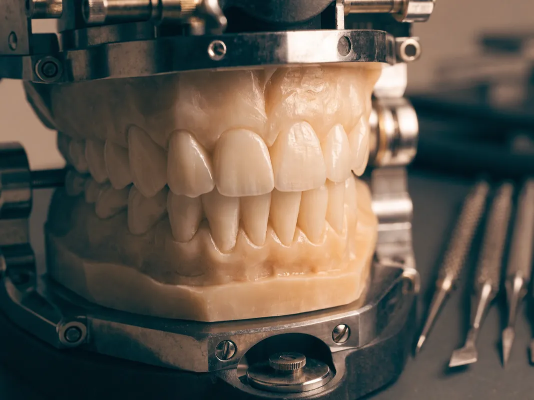
A reduction guide based on a diagnostic design may help the clinician prepare relative to the intended final surface rather than remove a uniform amount blindly. Before and after photographs, scans or impressions can document the baseline and preparation. The patient should understand which surfaces are altered and where dentine exposure is anticipated.
“Ninety per cent of the tooth remains” is not a meaningful consent statement without a measured design. No-preparation language also needs scrutiny: a restoration can be additive and still create irreversible changes through bonding, margin placement or future replacement.
Mock-up, trial, provisional and final restoration are different
These terms should not be blurred:
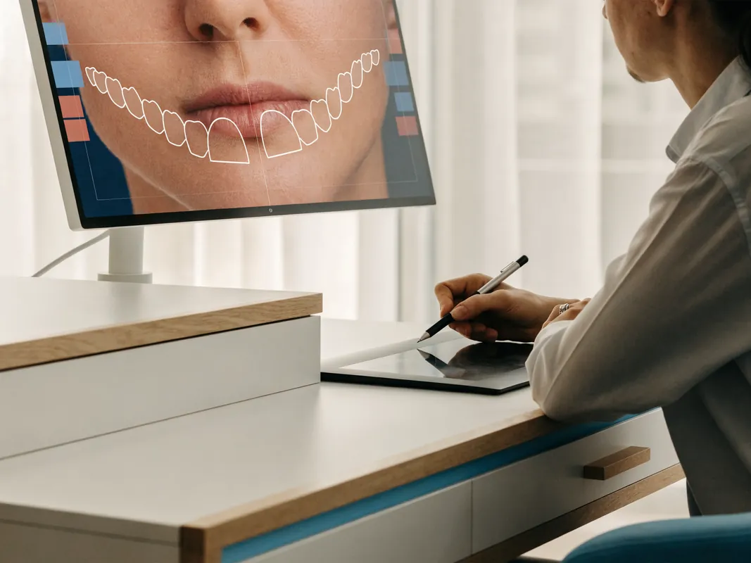
- a digital design is a visual planning proposal;
- a mock-up is a reversible or provisional representation placed over teeth to test shape and sometimes colour broadly;
- a preparation guide helps relate reduction to the proposed final contour;
- a provisional restoration protects prepared teeth and tests appearance, speech, function and tissue response during the laboratory phase;
- a try-in previews certain aspects of the fabricated restoration before definitive bonding, subject to the limits of try-in materials and lighting;
- the final restoration is accepted and bonded only after clinical checks and valid consent.
A mock-up cannot reproduce the final optical interaction perfectly, especially over a dark substrate. A digital design cannot show translucency reliably. A try-in paste may change apparent shade but does not guarantee the bonded result under every light. These limitations should be stated before preparation.
The provisional can test tooth length, contour, speech, lip support, contact and bite, but its material and opacity differ from the final restoration. It must allow gum health and cleaning. Problems should be corrected rather than dismissed because the final material will be different.
Shade selection needs a target and a controlled method
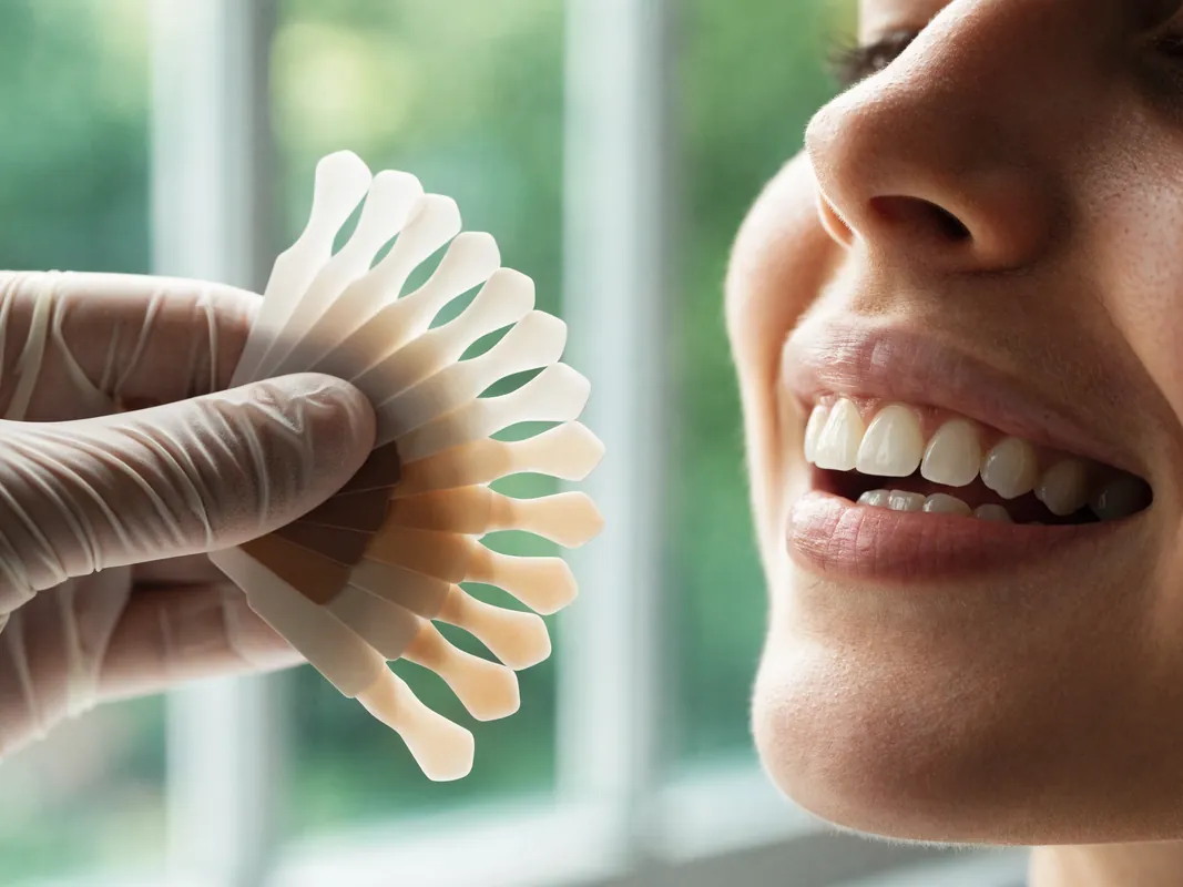
Shade is more than a tab code. Value, chroma, hue, translucency, fluorescence, texture, lustre and the colour of neighbouring teeth affect perception. Bands can vary from cervical to incisal areas, and whitening may change regions differently. The desired target may be a natural match, a modestly lighter smile or a substantially different aesthetic.
Shade communication may use visual guides, calibrated photography, spectrophotometry or other digital tools. Systematic reviews suggest digital approaches can improve consistency in some settings, but ambient light, device calibration, operator technique, dehydration and study quality matter. No instrument eliminates subjective acceptance.
Record:
- baseline shade by tooth and zone;
- substrate or preparation shade if indirect restorations are made;
- whitening history and stabilised post-whitening record where relevant;
- shade guides or device used;
- lighting and photographic calibration;
- proposed restorative material and opacity;
- patient-approved target and known limits;
- laboratory instructions and returned shade records;
- any remanufacture decision and reason.
The patient should view the trial in suitable conditions and understand that home, office and daylight can produce metameric differences.
Function and gum health can veto an aesthetic design
Adding length, width or facial volume can change speech, lip closure, guidance, contacts and cleaning. Deep bite, edge-to-edge contact, crossbite, tooth wear and parafunction can affect composite and ceramic choices. The dentist should test the design functionally and explain any occlusal adjustment, protective appliance or alternative.
A night guard may be considered for selected patients but does not guarantee against fracture or debonding. It needs fit, cleaning and review. Existing jaw pain or temporomandibular symptoms require a separate baseline; veneers cannot be promised to cure them.
Margins and contours should allow plaque control and periodontal assessment. Inflamed gums can recede after health improves, changing the visible margin. A provisional that traps plaque can distort the tissue before final bonding. The patient should demonstrate cleaning with the proposed shape.
Consent must include the aesthetic limits
GDC Principle 3 describes consent as a continuing process that includes relevant options, risks, benefits and costs, time for questions and the patient’s right to change their mind. For intrinsic discolouration, consent should explicitly reject guaranteed masking and a “one possible alternative after clinical assessment” narrative.
Discuss possible outcomes such as:
- partial rather than complete whitening response;
- residual bands or uneven colour;
- sensitivity or gum irritation during whitening;
- an existing restoration becoming mismatched;
- incomplete masking through composite or ceramic;
- a restoration appearing more opaque or flatter than desired;
- a visible transition between treated and untreated teeth;
- a difference under daylight, indoor light and photography;
- over-contour if masking is attempted without enough space;
- dentine exposure, sensitivity or pulp risk from preparation;
- gum recession or a visible margin;
- fracture, chipping, debonding, wear, stain or caries;
- need for repair, remanufacture, endodontic treatment or replacement;
- future whitening or natural tooth change affecting the match;
- inability to meet the desired shade without unacceptable tissue removal.
The patient must understand which elements are irreversible. Signing a form after tooth preparation is not a substitute for consent before preparation. If the masking strategy or number of teeth changes after a trial, explain the new trade-offs and obtain renewed consent.
The right to stop or accept a less dramatic result
Patients can decide that partial whitening is enough, choose composite instead of ceramic, treat fewer teeth, postpone, or stop after a diagnostic phase. A clinician should not frame acceptance of natural variation as treatment failure. Where the requested shade would demand excessive reduction or opacity, the responsible recommendation may be a more modest target or no indirect restoration.
Event dates, travel and deposits should not override this right. A patient who dislikes the mock-up or cannot accept the stated limits should have space to reconsider before irreversible work.
Laboratory and material records make future care possible
GDC Principle 4 supports complete records including clinical findings, radiographs, photographs, models or scans, consent, laboratory prescriptions, statements of conformity and referral communications where applicable. For a masking case, records should include:
- diagnosis and differential diagnosis;
- baseline tooth and gum health;
- whitening product and protocol if prescribed;
- sensitivity and response reviews;
- baseline, post-whitening and preparation shade records;
- preoperative photographs and diagnostic design;
- preparation scans or impressions;
- composite or ceramic material identity;
- cement, adhesive and surface-treatment records where relevant;
- laboratory name, prescription and manufacture location;
- lot or batch information where retained by the provider;
- final shade, design and occlusion;
- complications, repairs and maintenance instructions.
The MHRA guidance on custom-made devices explains documentation and responsibilities for relevant devices in Great Britain. Treatment abroad may fall under another regulatory route, but the patient still needs intelligible material and laboratory records for local repair and future replacement.
Avoid vague terms such as “premium ceramic” or “Swiss quality.” They do not identify the material, laboratory or responsible provider.
Demand an itemised written quote
A single “smile package” total hides the clinical decisions. Ask the legal provider for an itemised quote separating:
- examination, diagnosis and specialist opinions;
- photographs, scans, shade analysis and justified radiographs;
- cleaning and periodontal care;
- external or internal whitening, reviews and replacement trays or products;
- orthodontic assessment or treatment if relevant;
- composite trial, repair or veneers by tooth;
- indirect veneer or crown by tooth;
- mock-up, preparation guide and provisional restorations;
- material, laboratory, try-in and remanufacture stages;
- bonding, occlusal adjustment and protective appliance if prescribed;
- planned reviews, maintenance and records;
- treatment of complications or a changed plan;
- cancellation, postponement, refund and complaint terms;
- travel, accommodation and transfers as separate non-clinical items if purchased.
The quote should state what is excluded and what remains provisional until examination, whitening response, preparation or trial. Ask what happens financially if fewer teeth are treated, whitening meets the goal, composite replaces ceramic, a veneer plan becomes structurally inappropriate, a crown is proposed, the shade is rejected before bonding or the laboratory must remake a restoration.
“Free remedial treatment” can still leave flights, accommodation, time off work and local dentistry unpaid. Read the contract rather than assuming a warranty covers biology, subjective appearance or travel.
Remote reviews cannot finalise diagnosis or masking
A remote enquiry can collect medical and medication history, childhood exposure information, photographs, dental records, radiographs and goals. It can identify missing information and explain broad options. It cannot clean the teeth, distinguish every surface and intrinsic cause, test pulp, inspect cracks, assess enamel, measure gum health, check occlusion, verify lighting or determine preparation shade.
Any remote plan and quote should be provisional. Before sharing health data, identify the legal recipient, privacy notice, secure channel, purpose, access and retention. Compressed social-media photographs are poor shade records and may exaggerate or hide banding.
At the in-person visit, the dentist must confirm diagnosis, health, options, preparation, material strategy, risks and quote. A change requires explanation and continuing consent. The patient should not feel obliged to add teeth or accept crowns because travel is already booked.
Cross-border travel must follow clinical gates
The GDC patient guidance on dental care abroad and the NHS treatment-abroad checklist recommend verifying providers and qualifications, understanding procedures and costs, planning aftercare, checking complaint routes and considering insurance and return travel. These principles matter when a multi-tooth aesthetic plan depends on laboratory stages and subjective acceptance.
Before booking, obtain:
- legal provider name and clinic address;
- named treating dentist and registration route;
- laboratory name and where work is made;
- provisional sequence and what could change it;
- clinically required attendance with contingency for review or remanufacture;
- urgent contact and local aftercare route;
- insurance terms and exclusions;
- record-transfer plan;
- cancellation and delay terms;
- travel, hotel and transfer details separately from clinical care.
Do not let a return flight become a bonding deadline. If the trial is unacceptable, gums are inflamed, a tooth is symptomatic, the laboratory needs revision or the dentist recommends postponement, the itinerary should change rather than force irreversible completion.
Local aftercare is part of the initial decision
Identify a local dentist before extensive care abroad. Ask whether they are willing to review, clean and repair the proposed composite or ceramic system and what records they need. Do not assume every practice will manage another provider’s work immediately or can reproduce an undocumented shade and material.
Maintenance may include caries and periodontal review, margin inspection, plaque control, occlusion, sensitivity, cracks, chipping, debonding, stain, wear, gum recession and protective-appliance review. Natural teeth and restorations can change differently over time. Professional cleaning methods must be compatible with the materials.
Remote photographs can support communication but do not replace necessary clinical examination. A colour difference visible only under certain light may still require direct evaluation and comparable records.
Red flags before treatment
Pause and seek independent advice if you encounter:
- diagnosis of tetracycline staining from a filtered photograph alone;
- the claim that whitening never changes tetracycline-associated colour;
- a veneer or crown described as the only one possible alternative after clinical assessment;
- a severity grade used as a fixed prescription;
- composite dismissed without a case-specific explanation;
- no treatment, cleaning, whitening, orthodontic or combination options omitted;
- a fixed preparation depth or percentage of tooth “preserved” promised online;
- a material brand described as universally best;
- guaranteed complete masking, “real teeth” appearance or a life-changing result;
- a fixed number of upper and lower veneers sold as standard;
- healthy teeth proposed for crowns solely to create opacity;
- digital smile design presented as the final result;
- no mock-up, trial, provisional or change-of-plan pathway;
- no named dentist, legal provider or laboratory;
- consent managed only by sales staff;
- no material, shade, laboratory or device records;
- a fixed multi-day package that leaves no room for gum health or remanufacture;
- an expiring discount tied to same-day consent;
- no local maintenance or complaint route.
Red flags during whitening or after restorative treatment
Follow individual instructions and contact the responsible clinician for persistent or worsening sensitivity, gum burns or ulceration, severe pain, swelling, fracture, a loose restoration, a sudden bite change, inability to clean, bleeding that does not settle or another concerning change. Stop unsupervised products if tissue injury occurs and seek professional assessment.
Difficulty breathing or swallowing, rapidly increasing facial or neck swelling, uncontrolled bleeding, collapse or another medical emergency requires emergency care. The NHS urgent dental guidance explains how to obtain urgent dental help in the UK. Do not wait for an overseas message when local emergency assessment is needed.
Questions to ask the named dentist and laboratory
Use this list before consent:
- What evidence supports tetracycline-associated discolouration?
- Which alternative diagnoses were considered?
- Is the colour extrinsic, intrinsic, internalised or mixed?
- Are any teeth non-vital, carious, cracked or structurally compromised?
- What gum or functional problems need treatment first?
- Is no treatment or monitoring reasonable?
- What can professional cleaning change?
- Is supervised external whitening suitable?
- What response is uncertain, and what is the stopping rule?
- Does one tooth need a separate internal-whitening assessment?
- Could whitening reduce the number or opacity of restorations?
- Is direct composite a reasonable additive option?
- Would orthodontics address position or spacing first?
- Why is a veneer proposed for each named tooth?
- What structural finding would justify a crown?
- How much enamel can be preserved?
- What substrate colour will the laboratory receive?
- How will opacity and translucency be balanced?
- Which material and cement system are proposed, and why?
- Can a mock-up or composite trial test the goal?
- What can the provisional reveal?
- What can and cannot be judged at try-in?
- How are lighting and dehydration controlled during shade selection?
- What residual banding, mismatch or opacity cannot be ruled out?
- How will function and gum health be checked?
- What material, laboratory and shade records will I receive?
- What exactly is included and excluded in the itemised quote?
- What happens if I reject the shade before bonding?
- Who provides local aftercare and repairs?
- Can I take the records for an independent second opinion?
A patient decision checklist
Before irreversible treatment, confirm:
- The cause of discolouration is diagnosed rather than assumed.
- Oral disease, gum health and function are stabilised.
- My goals distinguish colour from shape, alignment and gum concerns.
- I have discussed no treatment, cleaning, whitening, composite, orthodontic, veneers and crowns where relevant.
- I understand whitening may help without completely correcting the pattern.
- I understand existing restorations will not whiten like natural teeth.
- I understand enamel preservation and future restoration cycles.
- The mock-up, provisional, try-in and final stages are clearly different.
- I understand complete masking and a specific shade are not guaranteed.
- The number of teeth follows my smile and decision, not a package count.
- I know the dentist, legal provider and laboratory.
- I will receive material, shade and device records.
- I have an itemised quote and change-of-plan terms.
- I have local maintenance and urgent-care routes.
- I can pause, seek a second opinion or decline treatment.
Evidence and further reading
These sources inform the decision framework. They do not diagnose an individual, prescribe a whitening regimen or guarantee a restorative result.
- American Dental Association, Whitening, accessed 29 August 2026: intrinsic and extrinsic discolouration, whitening mechanisms, safety, sensitivity and restoration limits. https://www.ada.org/resources/ada-library/oral-health-topics/whitening
- General Dental Council, Tooth whitening and illegal practice, accessed 29 August 2026: tooth whitening as dentistry and lawful professional provision in the UK. https://www.gdc-uk.org/standards-guidance/information-for-patients-public/tooth-whitening-and-illegal-practice
- NHS, Teeth whitening, accessed 29 August 2026: assessment, provider safety and risks of unregulated treatment. https://www.nhs.uk/tests-and-treatments/teeth-whitening/
- Randomised controlled trial of home bleaching in tetracycline-stained teeth, accessed 29 August 2026: measurable colour improvement in the study population and response uncertainty. https://pubmed.ncbi.nlm.nih.gov/28478214/
- Clinical trial of extended at-home bleaching for tetracycline-stained teeth, accessed 29 August 2026: studied colour change and sensitivity, not a universal prescription. https://pubmed.ncbi.nlm.nih.gov/12666888/
- Review of intrinsic, extrinsic and internalised tooth discolouration, accessed 29 August 2026: differential diagnosis and varied causes. https://pubmed.ncbi.nlm.nih.gov/16262034/
- Review of internal tooth whitening, accessed 29 August 2026: a distinct conservative pathway for selected non-vital discolouration. https://pubmed.ncbi.nlm.nih.gov/38753915/
- Clinical case report on conservative composite management of generalised tetracycline staining, accessed 29 August 2026: composite as a possible selected alternative with limited evidence. https://pubmed.ncbi.nlm.nih.gov/33339923/
- Systematic review of experimental masking methods, accessed 29 August 2026: substrate, material, thickness, pigment and cement variables and evidence limits. https://pubmed.ncbi.nlm.nih.gov/34085377/
- Systematic review of masking by manufactured ceramic restorations, accessed 29 August 2026: darker substrates, opacity, thickness and layering trade-offs, primarily laboratory evidence. https://pubmed.ncbi.nlm.nih.gov/39776151/
- Systematic review of veneer bonding to different tooth substrates, accessed 29 August 2026: importance of enamel preservation and limits of in-vitro evidence. https://pubmed.ncbi.nlm.nih.gov/39207840/
- Systematic review of porcelain laminate veneer survival and complications, accessed 29 August 2026: fractures, debonding and heterogeneous clinical evidence. https://pubmed.ncbi.nlm.nih.gov/33807504/
- Systematic review of digital dental shade matching, accessed 29 August 2026: lighting, devices, photography and methodological limitations. https://pubmed.ncbi.nlm.nih.gov/37999014/
- European Federation of Periodontology S3 guideline on peri-implant diseases, published 2023 and accessed 29 August 2026: disease control, cleanable design and supportive care principles. https://pubmed.ncbi.nlm.nih.gov/37271498/
- General Dental Council Principle 3, Obtain valid consent, accessed 29 August 2026: options, risks, benefits, costs, time and continuing consent. https://standards.gdc-uk.org/pages/principle3/principle3
- General Dental Council Principle 4, Maintain and protect patients’ information, accessed 29 August 2026: complete clinical, photographic and laboratory records. https://standards.gdc-uk.org/pages/principle4/principle4
- General Dental Council Principle 7, Work with colleagues in patients’ interests, accessed 29 August 2026: competence, collaboration and referral. https://standards.gdc-uk.org/pages/principle7/principle7
- Medicines and Healthcare products Regulatory Agency, custom-made medical devices in Great Britain, updated 20 July 2026 and accessed 29 August 2026: documentation and responsibilities for relevant devices. https://www.gov.uk/government/publications/custom-made-medical-devices/custom-made-devices-in-great-britain
- General Dental Council, Going abroad for dental treatment, accessed 29 August 2026: provider verification, records, costs, aftercare and complaints. https://www.gdc-uk.org/standards-guidance/information-for-patients-public/going-abroad-for-dental-treatment
- NHS, Treatment abroad checklist, accessed 29 August 2026: travel, insurance, follow-up and contingency questions. https://www.nhs.uk/using-the-nhs/healthcare-abroad/going-abroad-for-treatment/treatment-abroad-checklist/
- NHS, How to find an NHS dentist in an emergency, accessed 29 August 2026: urgent dental access and emergency warning signs. https://www.nhs.uk/nhs-services/dentists/how-to-find-an-nhs-dentist-in-an-emergency/
The evidence supports diagnosis, conservative sequencing, individual whitening assessment, enamel preservation and an honest masking trial. It does not support the claims that whitening can never change tetracycline-associated colour, that veneers are the only one possible alternative after clinical assessment, or that any material can guarantee complete masking.