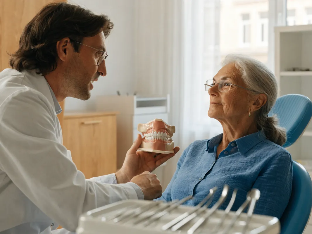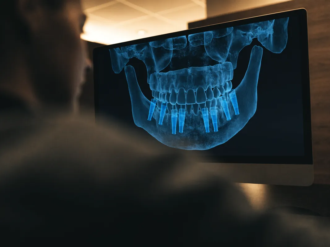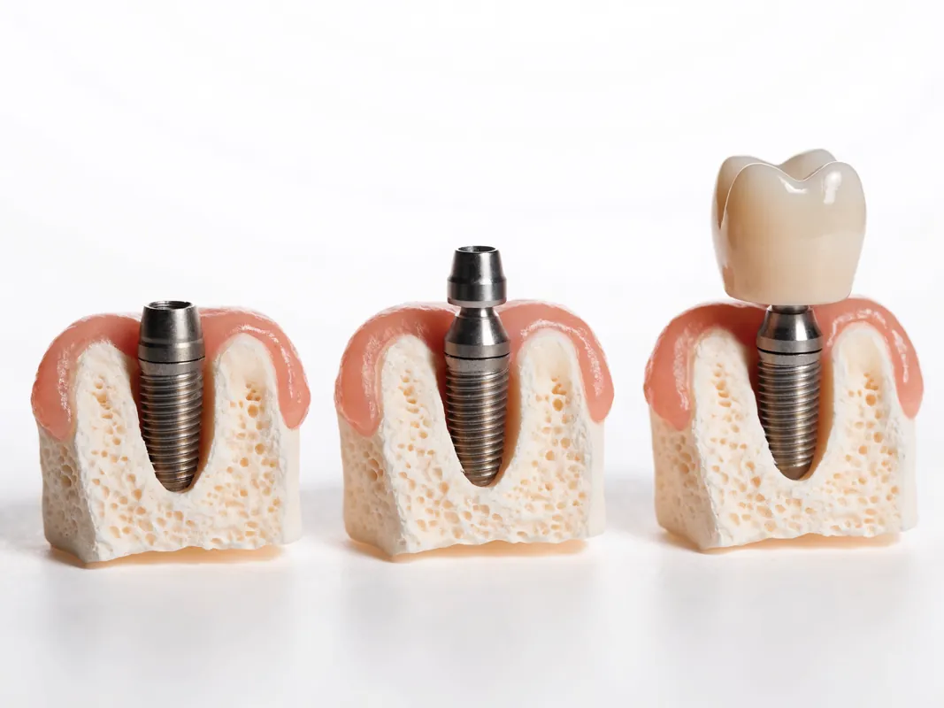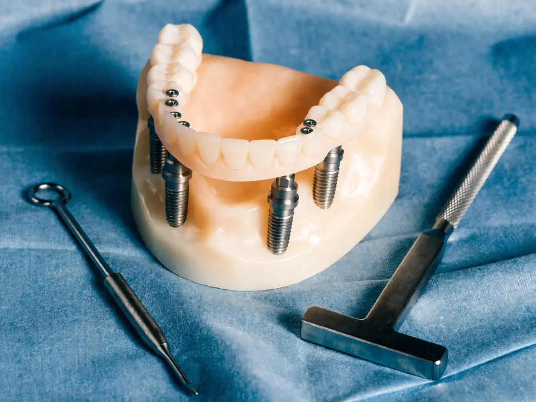
This guide uses six months as an orientation point for a structured review of a fixed full-arch implant restoration. It is not a universal appointment interval, a biological finish line or proof that treatment has succeeded. The responsible clinician and the local professional who examines the mouth must set review timing from the operation, restoration, medical history, periodontal history, symptoms, home-care access and findings. A person may need assessment before this point, at this point, later, or on a different sequence entirely.
The page is written for people who may hear the commercial term All-on-4, but the maintenance questions also apply to many fixed complete-arch implant restorations. A label does not reveal the number and position of implants actually present, the connection system, the restoration material, whether the bridge is provisional or definitive, how it is retained, or what maintenance access it permits. Begin with the patient-specific record rather than assuming that every full-arch bridge follows one protocol.
This is general information, not a remote examination. It cannot decide whether tissues are healthy, whether an implant is stable, whether a bridge should be removed, whether an image is justified, or when an individual should progress to another restorative stage. New or worsening symptoms override a calendar checklist. The clinician who assesses the person owns those decisions.
For broader implant recovery questions, use the phase-and-symptom implant aftercare guide. The All-on-4 service overview explains the treatment category. This page owns the maintenance-review intent: what to observe, what a professional may need to examine, what records should travel with the patient, and how to leave the review with an accountable next step.
What the six-month label means
Six months is a useful reminder to stop and reconcile the clinical, mechanical and practical record. It is not a rule that every patient must wait until six months or attend at exactly six months. The date may fall during provisional service, after a definitive bridge has been fitted, after an earlier complication, or long after a local professional has already begun supportive care. The relevant question is not whether the calendar was obeyed. It is whether the current status has been assessed at the interval selected for this person.
The evidence does not support turning a single interval into a universal website prescription. A systematic review specifically examining supportive care for full-arch implant prostheses found limited studies and no basis for drawing one precise frequency for prosthesis removal or maintenance. Its authors discussed possible intervals, but also said care should be tailored to the patient's risk profile and characteristics. Read the [systematic review abstract](https://pubmed.ncbi.nlm.nih.gov/38501398/) as evidence of uncertainty, not as permission to impose its suggested pattern on every mouth.
A written maintenance plan should therefore state who selected the interval, which findings informed it, what should trigger earlier contact, and who is available locally. If the plan simply says “six-month check” without naming the responsible professional, examination scope or escalation route, ask for those missing details.
First identify the restoration you actually have
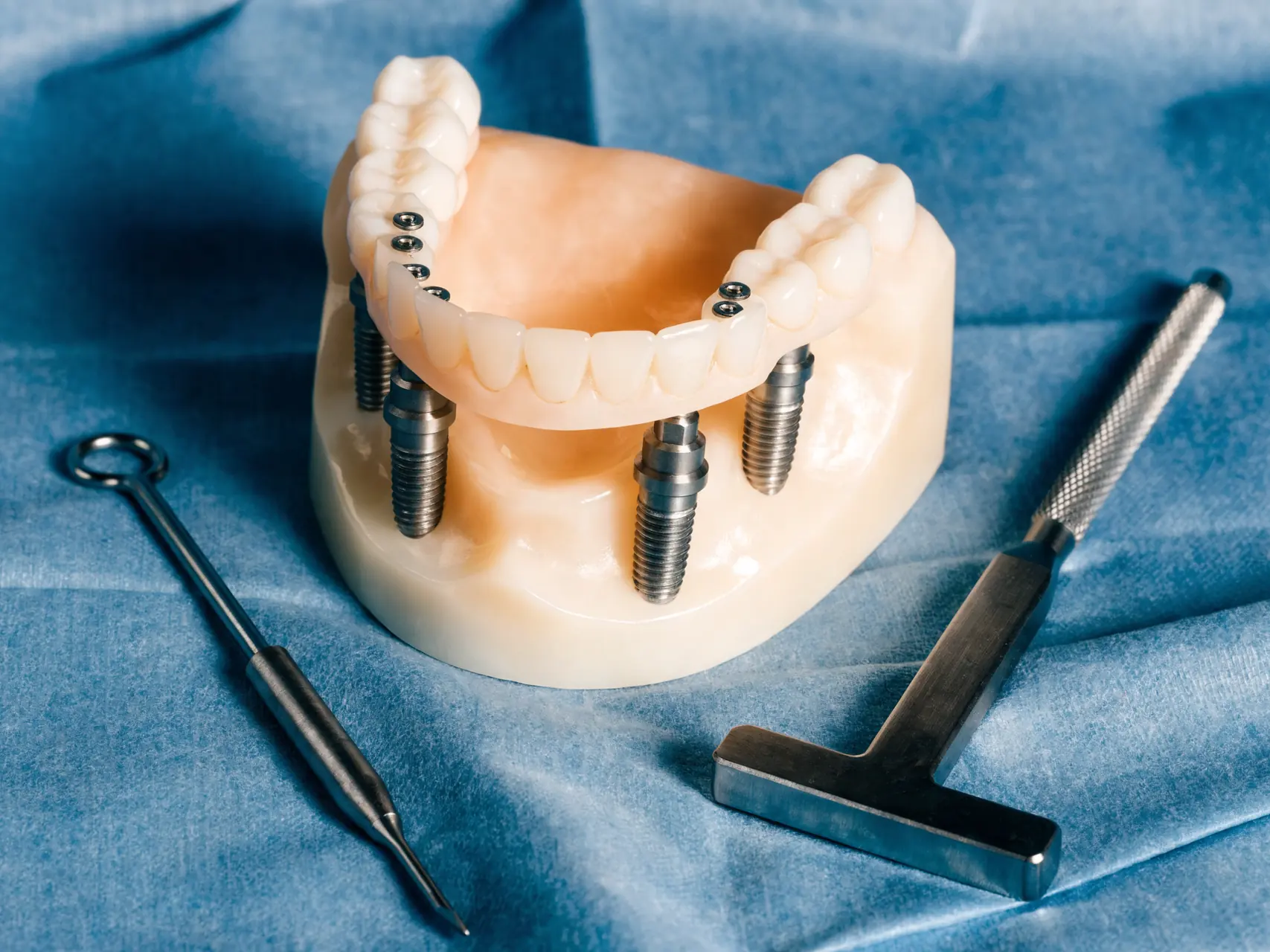
Before using any checklist, identify the current prosthetic state. “Fixed teeth” can refer to a provisional bridge fitted near surgery, a longer-term interim restoration used while tissues and function are assessed, or a definitive restoration accepted after clinical and laboratory stages. A bridge may be screw-retained, cement-retained or use another connection arrangement. Its framework and visible tooth material may be different materials. Access channels may be sealed. The underside contour may be designed for a particular cleaning route. None of this can be reconstructed reliably from colour or appearance alone.
Ask for a written statement answering these questions:
- Is this restoration provisional, interim or definitive?
- Which arch and which implants support it?
- Is it retained at implant level or abutment level, and by what method?
- What framework and veneering or tooth materials were used?
- Which components can be replaced independently?
- Which hygiene aids were selected for the actual underside and tissue contours?
- May a local professional remove it, and which instruments and parts would be required?
- Which clinical or laboratory stage remains outstanding?
Do not treat the words provisional and definitive as quality grades. A provisional restoration may be deliberately used to evaluate healing, speech, appearance, cleanability and loading. A definitive restoration can still require maintenance, repair or adaptation. The status matters because expectations, loading instructions, component records and decisions about removal may differ.
Assign responsibility before the review
A full-arch review can involve more than one professional, but responsibility must not dissolve between them. The original surgical clinician may hold the operative information. A restorative clinician may own the bridge design and occlusion. A hygienist or periodontal professional may provide supportive care within their scope. A local dentist may examine symptoms and coordinate urgent treatment. A laboratory may identify materials or fabricate a repair but cannot diagnose tissue disease. A facilitator or travel coordinator cannot replace clinical accountability.
Write a responsibility map before the appointment:
- Who performs the clinical examination?
- Who evaluates peri-implant tissues and periodontal risk?
- Who checks the bridge, components and bite?
- Who decides whether imaging is justified?
- Who can access or remove the restoration if clinically indicated?
- Who records components altered during the visit?
- Who receives an urgent message and who can provide face-to-face care?
- Who explains the next review interval and its reasons?
If the original treatment occurred abroad, identify a local dentist before a problem develops. The returning-home after dental tourism guide explains handover and local-care boundaries in detail. A local professional is not automatically responsible for treatment performed elsewhere and may lack the system, instruments, records or competence needed for a particular intervention. Ask rather than assume.
Build the pre-review record pack
The review is more useful when the professional can compare current findings with a reliable baseline. Collect documents rather than relying on a phone gallery or a brand name remembered from a conversation. The record pack should be organised by date and arch so that another professional can understand what happened without reverse-engineering the case.
Request, where applicable:
- final diagnosis and tooth or site chart;
- pre-treatment and post-treatment images with reports or clinical interpretation;
- surgical date, sites, procedures and named responsible clinicians;
- graft, membrane or sinus-related information if relevant;
- implant body, connection, diameter, length and lot or traceability identifiers;
- abutment, restorative base, fixation screw and other replaceable component references;
- torque or tightening record where clinically relevant and appropriate to release;
- provisional and definitive restoration dates and status;
- framework, tooth, veneering and repair materials;
- laboratory name, prescription, statement of conformity and design record where available;
- occlusal record, appliance information and documented adjustments;
- medicine and allergy history current at treatment and at review;
- personalised hygiene instruction and maintenance plan;
- prior symptoms, complications, repairs and components replaced;
- current clinical contact, emergency route, complaint procedure and commercial terms.
The [GDC records standard](https://standards.gdc-uk.org/pages/principle4/principle4) explains that complete patient records can include radiographs, consent forms, photographs, models, laboratory prescriptions, statements of conformity and referrals. That standard directly governs UK registrants; its record categories are also a useful continuity checklist when care crosses borders. It does not verify an overseas provider.
Prepare a dated symptom history
Do not reduce the review to “it feels fine” or “it hurts.” Prepare a short dated history covering change over time. Symptoms may arise from tissues, the restoration, components, muscles, joints, nerves, medicine effects, remaining teeth or another condition. The patient does not need to diagnose the cause. The aim is to give the examining professional usable information.
Record:
- the exact location and whether one or both arches are involved;
- when the change began and whether it is stable, intermittent or progressing;
- pain, pressure, tenderness, burning, altered sensation or numbness;
- bleeding during cleaning or spontaneous bleeding;
- swelling, discharge, bad taste or persistent odour;
- food trapping or a new area that cannot be cleaned;
- clicking, movement, fracture, roughness or a changed gap;
- altered bite, difficulty chewing, fatigue or avoidance of one side;
- speech change, lip or cheek biting and tongue irritation;
- trauma, heavy biting event or appliance damage;
- fever, systemic illness, medicine change or change in smoking or nicotine exposure;
- actions already taken and whether they changed the symptom.
A photograph can help document visible change, but it cannot test movement, probe tissues, inspect the entire fitting surface, assess the bite or replace appropriate imaging. Do not postpone face-to-face care because a remote image looks acceptable.
Urgent red flags come before maintenance
A routine review list is not an emergency triage tool. Seek urgent dental or medical assessment when symptoms indicate that waiting for the planned maintenance appointment may be unsafe. The appropriate route depends on location and severity. Use local emergency services for breathing or swallowing difficulty, major uncontrolled bleeding, collapse, severe systemic illness or rapidly spreading swelling rather than waiting for an overseas reply.
The [NHS dental abscess page](https://www.nhs.uk/conditions/dental-abscess/) describes urgent dental symptoms and emergency features including difficulty breathing, speaking or swallowing, substantial mouth swelling, eye involvement and severe limitation in opening the mouth. [NHS England urgent dental guidance](https://www.england.nhs.uk/long-read/clinical-guidance-unscheduled-urgent-and-non-urgent-dental-care/) distinguishes life-threatening, urgent and non-urgent presentations. Those routes apply in England; elsewhere use the equivalent local service.
Mechanical changes also need timely assessment. A bridge or component that is mobile, fractured, displaced or creating trauma should not be tested repeatedly by biting, pushed back with household tools, glued, filed or tightened by the patient. Stop using the affected area as instructed and obtain professional triage. A loose component may create biological, mechanical or airway concerns depending on the finding. Remote advice may help choose the route but cannot establish the cause.
Review comfort and function without assuming success
Function is broader than whether food can be chewed. Ask how the restoration behaves during speech, swallowing, smiling, light contact, firmer food and prolonged use. Compare current function with the earliest stable baseline rather than with a promotional expectation. A person may adapt to a gradual change and stop noticing it, so specific questions are more useful than a general satisfaction score.
Discuss:
- whether chewing has become one-sided;
- whether a particular area contacts first;
- whether food choices changed because of fear, discomfort or movement;
- whether speech changed recently or never stabilised;
- whether cheeks, lips or tongue are being bitten;
- whether fatigue, jaw pain or headache accompanies use;
- whether the restoration affects swallowing or creates gagging;
- whether an opposing denture, implant bridge or natural teeth have changed;
- whether the patient hears a click or feels flexing;
- whether the bridge appearance, lip support or tooth position creates a new functional problem.
None of these answers proves implant failure or prosthesis failure. They help the clinician decide what to examine. A restoration can remain in place while a repairable mechanical issue, tissue inflammation or occlusal change develops. Conversely, unfamiliar sensation does not automatically mean a component is failing.

Check the prosthesis as a system
The visible bridge is only one part of a connected system. Maintenance should consider the prosthetic teeth or ceramic, framework, fixation elements, access-channel material, abutments or bases, implant connections and implant bodies. Different parts can show wear or change for different reasons. The patient should not be expected to identify which layer is responsible.
Report roughness, chipping, cracks, staining that does not clean, an open access channel, a lost filling over a screw channel, a changed seam, food trapping, odour, clicking or movement. The professional may inspect for wear, fracture, loss of retention, component loosening, material separation, cleansability and whether any surface is injuring tissue. The method depends on the design and findings.
Evidence reviews show why “the bridge survived” is an incomplete outcome. A [systematic review of screw- and cement-retained full-arch restorations](https://pubmed.ncbi.nlm.nih.gov/32186285/) found different biological and technical complication patterns between retention approaches. This does not rank one design for an individual or predict an event. It supports recording the actual retention method and watching both biological and mechanical domains.
Do not allow a non-clinical label such as zirconia, acrylic, hybrid or permanent to substitute for the material and component map. The precise design influences how the restoration can be inspected, polished, repaired, removed and cleaned.
Occlusion and parafunction require clinical assessment
Occlusion means the way the upper and lower teeth or restorations contact during closure and movement. A patient can notice a first contact, slide, new impact, repeated cheek biting or fatigue, but cannot reliably measure the whole system. The clinician may need to assess static and moving contacts, opposing teeth or prostheses, wear, mobility, muscle and joint symptoms, component history and whether the bridge design distributes load as intended.
Do not self-adjust the bridge with a file, abrasive strip or rotary tool. Do not add a shop-bought guard over a full-arch restoration without clinical assessment. An appliance can alter contacts or fail to fit the actual bridge and opposing arch. If an appliance was prescribed, bring it to the review. Report whether it still seats fully, whether it is worn, whether it causes pressure, and whether symptoms changed while using it.
Grinding and clenching history should be documented rather than treated as a moral failure or a simple yes/no question. Sleep-related activity, daytime bracing, stress, medicines, pain and other factors may be relevant. A protective strategy may include education, monitoring, appliance review, repair planning or another clinical pathway, but no single measure guarantees protection.
The review output should record the occlusal findings and any change made. If a component was accessed or tightened, document what was found and which part was altered. “Bite checked” is too vague for future continuity.
Inspect cleanability, not only cleanliness
Plaque at a review may reflect technique, access, dexterity, vision, motivation, surface roughness, restoration contour, tissue change, pain, disability or inadequate instruction. A useful assessment asks both whether deposits are present and whether the person can physically reach the relevant surfaces without causing injury.
Ask the patient to demonstrate the actual home routine with their aids. The professional can observe which surfaces are missed, whether an aid enters the intended space, whether force is excessive, and whether a handle or adapted grip would help. Advice should be matched to the bridge design, tissue condition and abilities. A generic bag of products is not an individual hygiene plan.
A small crossover study of nine people with fixed full-arch prostheses compared powered tools and manual brushing and found that cleaning performance varied by method and by baseline ability. Read the [primary study](https://pubmed.ncbi.nlm.nih.gov/30911853/) for its small sample and design limitations. It does not establish one mandatory device or prove that a powered product cleans every surface.
A separate [split-mouth randomised trial](https://pubmed.ncbi.nlm.nih.gov/37171039/) found that prosthetic tissue-contact design affected plaque accumulation in its study setting. The broader lesson is that contour can affect access. When an area remains inaccessible despite coached technique, ask whether professional maintenance, prosthetic modification or another clinical response is indicated rather than blaming the patient.
Home care should be demonstrated and personalised
Home care usually needs more than polishing the visible front surfaces. The actual plan may include brushing accessible bridge surfaces, cleaning around transition zones, using an appropriately sized interdental aid or threader, and reaching the underside where the design permits. The professional should select tools after seeing the restoration and should demonstrate angle, route, pressure and frequency for this individual.
The [EFP prevention guideline summary](https://www.efp.org/fileadmin/uploads/efp/Documents/Other_publications/Clinical_guidelines/peri-implantitis-guideline-01-prevention.pdf) recommends individually tailored oral-hygiene instruction and demonstration to an oral healthcare professional, with periodic reinforcement. It also supports patient-centred supportive peri-implant care. This is a professional guideline, not an instruction to copy a product list from a website.
At the review, ask:
- Can the patient identify every intended cleaning route?
- Does the selected aid pass without shredding, locking or injuring tissue?
- Can the patient see or feel the correct path?
- Is bleeding persistent, increasing or localised?
- Does pain prevent cleaning?
- Is there an inaccessible surface under the bridge?
- Are manual dexterity, arthritis, tremor, visual limitation or caregiving needs relevant?
- Is a professional demonstration documented?
- When should a change prompt earlier examination?
If tissue bleeds, do not stop all cleaning indefinitely or intensify force blindly. Arrange assessment and obtain tailored instruction. Bleeding can have more than one explanation, and the website cannot identify it.
Know the limits of home devices and rinses
A water irrigator, powered brush, single-tufted brush, interdental brush, floss threader or rinse may be useful in a particular plan, but no device performs a clinical examination or professional debridement. The evidence for one tool may come from small samples, different prostheses or short follow-up. Product marketing often makes broader claims than the study supports.
Do not force an aid into a space, use metal household objects, scrape components, place corrosive or abrasive substances under the bridge, or use concentrated chemicals without professional direction. An aid that catches, frays, locks or causes repeated trauma needs review. Do not assume that a stronger jet or larger brush solves an access problem created by contour or tissue change.
Mouthwash is not a substitute for mechanical plaque control or diagnosis. A clinician may recommend a product for a specific reason and duration, considering allergies, interactions, staining, taste disturbance, tissue condition and other factors. Follow the product instructions and the personal plan. Do not continue a medicated rinse indefinitely merely because it was part of early surgical instructions.
Home photography can track visible change but is limited by angle, light, focus and inaccessible surfaces. A clean-looking photograph does not show probing findings, mobility, occlusion or bone. Treat remote images as records for triage, not clearance.
Professional supportive peri-implant care
Professional maintenance should be based on current findings and individual risk. It may include review of medical and dental history, symptoms, plaque control, tissue appearance, probing where appropriate, bleeding or suppuration findings, restorative contour, implant and prosthetic stability, occlusion, remaining teeth, dentures and justified imaging. The exact instruments and methods must be compatible with the tissues, components and restoration.
The EFP S3 guideline on [prevention and treatment of peri-implant diseases](https://onlinelibrary.wiley.com/doi/full/10.1111/jcpe.13823) supports risk assessment, oral-hygiene instruction and supportive peri-implant care. The guidance is not a promise that maintenance prevents every complication, nor does it create the same interval for every patient. Risk can change with disease history, plaque control, smoking, diabetes, adherence, prosthetic access and new clinical findings.
Ask the professional to document what was assessed and what was done. “Cleaning completed” does not show which sites were accessible, whether the bridge was removed, which surfaces were debrided, whether tissue findings changed, or what advice followed. The record should be usable by the next professional.
Professional maintenance and repair are different tasks. A hygienist may identify a possible mechanical change but not undertake restorative intervention outside their scope. A dentist may need laboratory or specialist support. Define the referral route rather than assuming one appointment resolves every domain.
Peri-implant tissue assessment needs a baseline
Peri-implant tissues cannot be judged from colour alone. The professional may compare plaque, bleeding, suppuration, probing measurements where appropriate, tissue level, discomfort and radiographic bone levels when imaging is clinically justified. Interpretation is stronger when baseline records exist from the time the prosthesis was fitted or from a stable earlier review.
The [World Workshop consensus report](https://www.efp.org/fileadmin/uploads/efp/Documents/Campaigns/New_Classification/Reports/Consensus_report__Workgroup_4__Berglundh_et_al-2018-Journal_of_Clinical_Perontology.pdf) describes peri-implant mucositis and peri-implantitis as distinct clinical conditions and notes risk linked with severe periodontitis history, poor plaque control and absence of regular maintenance. The report is a classification and evidence consensus, not a remote diagnostic checklist.
A single number without context may mislead. Probe design, force, access, tissue anatomy, restoration contour and baseline differences can matter. A radiograph also requires consistent projection and clinical interpretation. Ask the clinician to explain whether a finding is new, whether it is compared with a baseline, what uncertainty remains and what response is recommended.
Do not call every bleeding site peri-implantitis, and do not call an absence of pain healthy. Tissue disease can exist without discomfort, while pain can arise from non-implant causes. Diagnosis belongs to the examining clinician.

Imaging is risk-based, not a calendar ritual
Six months does not automatically require a panoramic image, CBCT scan or any other fixed image set. The clinician should justify imaging from the question being asked, existing records, symptoms, examination findings and radiation-protection principles. New imaging should add information that can affect management.
Bring prior original images and reports where possible. Screenshots, compressed messaging images and photographs of films may lose detail or calibration. The reviewing professional should know when and why each image was made. If a new image is proposed, ask what clinical question it answers, whether a lower-dose or smaller-field option is suitable, and how it will be compared with the baseline.
CBCT is not a “comprehensive check” badge. It exposes the patient to ionising radiation and should have a clinical indication. Conversely, avoiding all imaging regardless of symptoms or findings is not a patient-centred rule. The decision is case-specific.
Keep the interpretation with the image. A future clinician needs more than the file: they need date, anatomy, projection or field, relevant report and clinical context. Record transfer must use a secure route appropriate to health information.
Prosthesis removal is not automatic
Some fixed full-arch restorations can be removed professionally; others may be harder to retrieve, and removal itself can introduce mechanical work, cost and risk. A six-month label does not mean that every bridge must be removed. The decision should follow symptoms, tissue access, maintenance findings, retention design, component history, clinician competence, manufacturer instructions and whether removal is likely to change care.
The systematic review on full-arch supportive care found insufficient evidence for one removal frequency. A [randomised crossover study](https://pubmed.ncbi.nlm.nih.gov/32938869/) of professional plaque removal with and without superstructure removal reported bacterial reduction in both study conditions. Its limited setting does not establish a universal approach. It shows why the clinical question should be defined instead of treating removal as a ritual.
If removal is proposed, ask:
- What finding or maintenance objective justifies it now?
- Which clinician will perform it and with which system-specific instruments?
- Are replacement fixation components or access materials available?
- What happens if a component is damaged or cannot be reused?
- How will the fitting surface and tissues be documented?
- What tightening and verification process follows refitting?
- Which component identifiers and changes will be added to the record?
- What costs and contingencies are stated in writing?
The patient must not attempt removal or tightening. A general screwdriver, adhesive or improvised tool can damage components, tissue or the restoration.
Provisional restoration status at the review
If the bridge is still provisional, the review should state why, what it is being used to evaluate, and which criteria govern the next step. Time elapsed alone does not make a provisional restoration definitive. The plan may be waiting for biological findings, tissue maturation, functional evaluation, laboratory stages, risk control, patient acceptance or another condition. The responsible clinician should explain the reason and uncertainty.
Document provisional material, reinforcement or framework, repair history, access design, loading instructions, occlusal changes, appearance and cleanability. Ask whether a fracture or repair changes the plan. A repaired provisional may still serve its purpose in one case and may require replacement or redesign in another. The website cannot decide.
The patient should understand what is not yet final: tooth arrangement, speech, shade, contour, underside access, bite, laboratory material, component selection or fit. A trial stage is useful only if observations are recorded and influence the next decision.
Do not accept pressure to move to a definitive restoration solely because travel was booked or a package deadline arrived. Equally, do not assume that leaving a provisional indefinitely is harmless. Ask for clinical reasons, alternatives, risks, expected maintenance and a written next-step owner.
Definitive restoration status at the review
Definitive means that a planned restorative stage has been accepted and delivered; it does not mean permanent, maintenance-free or guaranteed. The review should establish a stable baseline for future comparison. Record appearance, speech, contacts, cleansability, tissue findings, implant and component identifiers, retention method, materials and any appliance supplied.
Ask whether the bridge can be repaired locally, whether a laboratory design file or physical model exists, whether the material requires a specific protocol, and which parts remain available. A definitive restoration may later need polishing, access-channel replacement, screw or component work, tooth or ceramic repair, reline of an opposing prosthesis, occlusal adjustment or more extensive intervention. The possibility of maintenance should be discussed without predicting that one event will occur.
Acceptance should be informed. If the patient reports unresolved function, pain, hygiene access or appearance concerns, those should be examined and documented rather than concealed by the word final. The clinician should explain reasonable options, risks, costs and the consequence of monitoring or delaying action.
Store the definitive handover separately from promotional material. It is a clinical and technical record that should remain usable if the original business, clinician, laboratory or distributor later changes.
Create a component traceability map
Future serviceability depends on more than the implant brand. A full-arch restoration can include implant bodies, multi-unit or other abutments, restorative bases, cylinders, fixation screws, framework, prosthetic teeth or ceramic, access materials and an appliance. Parts may come from different sources or be replaced at different visits. Record them at component level.
The map should include manufacturer, product or family name, reference, lot or other traceability identifier where supplied, connection, dimensions where relevant, site and date placed or changed. It should distinguish original from compatible or third-party components without implying that a category is automatically suitable or unsuitable. Suitability and compatibility are professional decisions based on the exact system and applicable instructions.
The implant-system verification guide gives a detailed method for building this record. The [FDA unique device identification overview](https://www.fda.gov/medical-devices/unique-device-identification-system-udi-system/unique-device-identification-system-udi-system) explains the purpose of device identification in the United States regulatory context. UDI availability and legal requirements differ by device, market and date; absence of a UDI field does not allow the website to identify a product.
When a component is accessed or replaced, update the map. Do not overwrite the old entry. Future clinicians need the history: what remained, what changed, why it changed and which professional performed the intervention.
Record materials and laboratory information
A patient-facing record should name the restoration materials precisely enough to support future care. “Zirconia teeth,” “acrylic bridge” or “premium ceramic” may omit framework, veneering, bonding, repair and access-channel materials. Ask for the laboratory prescription and statement of conformity where applicable, plus the laboratory identity and relevant design record.
Material identity matters when a professional considers polishing, repair, bonding, adjustment or remake. A technique suitable for one surface may not suit another. Surface roughness, wear, fracture and staining need assessment rather than a household repair method.
If digital files exist, ask what they contain, who controls them, how long they are retained, and how a copy can be obtained. A scan alone may not include occlusion, verified fit, component identity or laboratory decisions. If a physical model, index or verification jig exists, record its status.
Do not infer quality from country labels, trade language or a laboratory photograph. Traceability tells future professionals what was used; it does not prove that the diagnosis, design or execution was appropriate. Keep clinical, technical and commercial questions separate.
Review medical, periodontal and behaviour changes
The maintenance plan should be updated when health or behaviour changes. Bring a current medicine list, allergies, diagnoses, hospital care, smoking or nicotine history, diabetes information if relevant, and any new condition affecting healing, bleeding, dry mouth, dexterity, immunity or attendance. Do not stop or alter prescribed medicine for a dental review without the relevant prescriber and dental clinician.
Periodontal history matters even when no natural teeth remain in the treated arch. Prior disease, plaque control and supportive-care attendance can inform risk assessment. Remaining natural teeth and the opposing arch also need care. A full-arch bridge does not isolate the implants from the rest of the mouth.
Smoking, vaping and nicotine exposure should be recorded neutrally and accurately. A clinician can discuss risk, support and alternatives without guaranteeing that cessation prevents a complication or treating continued exposure as the sole cause of every finding. Alcohol, nutrition, sleep and parafunction may also be relevant depending on the case.
Practical ability can change. Arthritis, tremor, visual impairment, cognitive change, reduced grip, caregiving arrangements or financial barriers may make the original home-care plan unworkable. Maintenance should adapt to the person, not merely repeat an instruction they cannot perform.
Local handover after treatment abroad
Cross-border treatment needs a local plan before the maintenance date arrives. Contact a local dentist, explain the exact restoration and ask what care they are willing and equipped to provide. Share the proposed record pack securely. Ask whether they can offer routine examination, supportive peri-implant care, urgent assessment, imaging when justified, prosthetic access or referral. One practice may accept some tasks and decline others.
The GDC's [guidance on dental treatment abroad](https://www.gdc-uk.org/standards-guidance/information-for-patients-public/going-abroad-for-dental-treatment) advises patients to investigate who provides treatment, aftercare, complications, additional travel, complaints and costs. It also suggests speaking to a dentist at home. The GDC does not regulate every overseas professional, and its page does not certify any foreign provider.
Give the local professional a direct clinical contact rather than only a sales or logistics number. Consent is needed for record sharing. Translation may be required, but translated summaries should remain linked to the original records and dates.
If no local professional can support the system, treat that as a serviceability and travel-planning issue. It does not automatically mean treatment has failed or that travel is required. It means the responsible clinicians should explain feasible options, urgency, risks, costs and what can safely wait for a planned review.

Remote review has strict limits
Video, photographs and messaging can help collect history, inspect a limited visible area, share records and decide whether face-to-face assessment is needed. They cannot palpate tissues, test mobility, probe sites, assess the full bite, inspect every underside surface, obtain new imaging, remove a restoration or distinguish every biological and mechanical cause.
A remote contact should therefore produce a triage output, not a declaration that everything is healthy. The output should state who reviewed the information, what information was available, what could not be assessed, the recommended urgency, the face-to-face route and symptoms that require escalation.
Do not accept “send a photo at six months” as the entire maintenance plan. Do not delay local urgent care while waiting for a reply across time zones. A remote clinician may remain responsible for advice they give, but the local examining clinician makes an independent decision for care they provide.
Protect health information. Confirm the recipient, legal entity, privacy notice, secure transfer method, retention policy and who can access the images. Avoid sending passports, medical records or identifiable facial images into an unverified group chat.
Travel for review is a separate decision
A clinical need for assessment does not automatically prove that international travel is required. First identify what must be done, who can do it locally, how urgent it is, what records and parts are needed, and whether waiting changes risk. Compare a local examination, specialist referral, planned return and other appropriate options.
If travel is considered, the itinerary must remain subordinate to clinical findings. Ask who pays if the plan changes, whether accommodation and transport are separately booked, what happens if an extended stay is required, and whether insurance addresses planned treatment or related complications. Do not assume that a marketing package covers remedial work.
The patient should also assess fitness to travel, medicines, accessibility, companion needs and emergency access. A dental professional assesses the clinical issue; an airline controls carriage; an insurer interprets its policy; immigration and customs authorities control entry and medicines. Those responsibilities should not be collapsed into one coordinator's assurance.
For detailed travel and handover planning, use the returning-home guide linked earlier. The maintenance checklist remains valid even when every review occurs locally.
Written complaint and remedial terms
Clinical aftercare exists whether or not a commercial warranty exists. A patient with urgent symptoms should receive appropriate triage and care while contract questions are documented separately. Do not delay examination until liability, reimbursement or travel payment is agreed.
Before treatment and again at handover, ask for the written contract and complaint procedure. The dental warranty and guarantee guide explains how to compare commercial wording without treating it as a clinical promise. At the review, ask:
- Which legal entity contracted for the treatment and restoration?
- Which clinician and facility provided each stage?
- What is covered, excluded and time-limited in the written terms?
- Are examination, diagnosis, local care, laboratory work, components, travel and accommodation treated separately?
- Who decides whether a problem meets a commercial condition?
- What evidence and maintenance records are required?
- Who pays for local assessment before any return decision?
- What happens if the original clinician, laboratory or component is unavailable?
- Which complaint route and regulator apply?
- Does signing a settlement affect access to records or independent care?
The [GDC complaints principle](https://standards.gdc-uk.org/pages/principle5/principle5) requires a clear and effective complaint procedure for UK registrants. Overseas routes differ. Verify the correct provider, regulator, jurisdiction and deadlines independently. This page makes no warranty, refund, repair, travel or outcome promise.
A structured six-month review checklist
Use the following list as an agenda with the examining professional, not as a self-certification form.
Current status
- [ ] The restoration is identified as provisional, interim or definitive.
- [ ] The actual implant and retention design is recorded.
- [ ] Outstanding clinical or laboratory stages are named.
- [ ] The responsible clinicians and local professional are identified.
Symptoms and urgent care
- [ ] A dated symptom history has been reviewed.
- [ ] Pain, swelling, bleeding, discharge, taste, odour and sensation changes are documented.
- [ ] Mobility, clicking, fracture, roughness and food trapping are documented.
- [ ] The patient knows which signs need emergency, urgent or routine care.
Function and prosthesis
- [ ] Chewing, speech, swallowing and tissue comfort have been discussed.
- [ ] Occlusion and opposing teeth or prostheses have been assessed as indicated.
- [ ] Wear, access channels, retention and visible material changes have been inspected.
- [ ] Any adjustment, access or component change is recorded precisely.
Tissues and hygiene
- [ ] Peri-implant and periodontal findings are compared with a useful baseline where available.
- [ ] The patient demonstrates the actual home-care method.
- [ ] Cleaning aids match the bridge contour, tissue condition and ability.
- [ ] Inaccessible surfaces have a professional plan rather than blame.
- [ ] Professional supportive care and its next interval are individually justified.
Records and continuity
- [ ] Images are clinically justified and kept with interpretation.
- [ ] Implant, abutment, fixation and restorative components are traceable.
- [ ] Materials and laboratory information are available.
- [ ] The local handover route and remote-review limits are written.
- [ ] Complaint and remedial terms remain separate from clinical advice.
Questions to ask during the appointment
Ask direct questions and request answers in ordinary language:
- What is the current clinical and prosthetic status of each arch?
- Which findings are new compared with the baseline?
- Which findings cannot be assessed today and why?
- Does any symptom need urgent investigation or treatment?
- Is the bridge provisional or definitive, and what remains outstanding?
- Can I reach every intended cleaning surface with my current tools?
- Please watch me demonstrate the cleaning method; what should change?
- Is professional debridement needed, and which surfaces can be accessed?
- Is prosthesis removal indicated now? What finding supports that decision?
- Is imaging justified? What question will it answer?
- What did the occlusal examination show?
- Are any components loose, worn, fractured, altered or unavailable?
- Has the component and material record been updated?
- What is the next review interval for me, and why?
- What specific change should trigger an earlier appointment?
- Which clinician owns each next action?
- Which parts of future care can be provided locally?
- What written costs and commercial terms apply to any proposed work?
The answer “come back in six months” is incomplete unless the clinician explains the current findings, reason for that interval and earlier escalation triggers.
What the review should produce
Leave with an updated record, not only verbal reassurance. The output should include the date, location, examining professional, medical-history update, symptoms, clinical findings, prosthetic status, hygiene assessment, tissue findings, occlusal findings, imaging and interpretation if used, professional maintenance performed, components accessed or replaced, materials used, advice demonstrated, warning signs, next interval and responsible contact.
If treatment is proposed, request diagnosis, reasonable alternatives, risks, expected benefits, uncertainty, costs and consequences of monitoring or no treatment. If a plan changes from provisional to definitive, the new consent and quotation should reflect the change. If no intervention is required, record the basis for monitoring and the signs that would reopen the decision.
Provide the updated record to the local professional and original clinicians with consent. Keep a patient copy in a durable format. Add rather than replace previous records so the component and symptom history remains visible.
Evidence limits and source-led interpretation
Full-arch implant research uses different prostheses, populations, maintenance methods, definitions and follow-up. A study may report that a restoration remained in function while still recording repairs or tissue treatment. Group results do not predict one person's outcome. Systematic reviews can inherit limitations and inconsistent definitions from their included studies.
The [review of peri-implant disease in full-arch restorations](https://pubmed.ncbi.nlm.nih.gov/33571325/) found substantial variation in reported disease estimates and methods. It supports careful assessment but does not diagnose an individual. The [framework-material systematic review](https://pubmed.ncbi.nlm.nih.gov/34204681/) describes material and design evidence with limitations; it does not prove that one advertised material is best for a particular person.
The [FDA dental implant patient information](https://www.fda.gov/medical-devices/dental-devices/dental-implants-what-you-should-know) explains that implant systems include more than the implant body and that risks and benefits require discussion. The FDA regulates within the United States; its page does not approve a provider or decide a treatment plan elsewhere.
Official and primary sources were checked on 29 August 2026. Guidance, pages, regulations and evidence can change, so check the current source and local rules when care is planned. The sources support risk-based maintenance, records, individual assessment and urgent-care boundaries. They do not endorse a clinic, schedule, bridge material, package or outcome.
Final decision frame
At the six-month orientation point, the useful outcome is not a pass or fail sticker. It is a clear state description and an accountable plan.
Continue the current plan only when the examining professional has assessed the relevant tissues, prosthesis, function and hygiene and has documented why monitoring and the selected interval are appropriate.
Modify maintenance or the restoration plan when access, technique, symptoms, tissue findings, occlusion, provisional status, component history or patient ability requires change. The modification should name the responsible clinician, risks, alternatives, costs and follow-up.
Escalate promptly when urgent biological or mechanical signs are present, when the bridge is mobile or causing injury, when spreading swelling or uncontrolled bleeding occurs, or when the patient becomes systemically unwell. Emergency symptoms take priority over contracts and travel plans.
Pause an irreversible or definitive step when diagnosis, consent, component identity, cleanability, local handover or responsibility remains unresolved. A booked trip, elapsed time or commercial deadline is not a clinical indication.
Six months can be a valuable prompt because it encourages reconciliation of symptoms, function, hygiene, tissues, occlusion, records, components and continuity. Its value comes from the quality of the assessment, not from the date itself.